Anticorps Polyclonal de lapin anti-HA tag
HA tag Polyclonal Antibody for WB, IF/ICC, FC (Intra), IP, ELISA
Hôte / Isotype
Lapin / IgG
Réactivité testée
Protéine recombinante et plus (4)
Applications
WB, IF/ICC, FC (Intra), IP, CoIP, ChIP, ELISA
Conjugaison
Non conjugué
N° de cat : 51064-2-AP
Synonymes
 at various dilutions.")

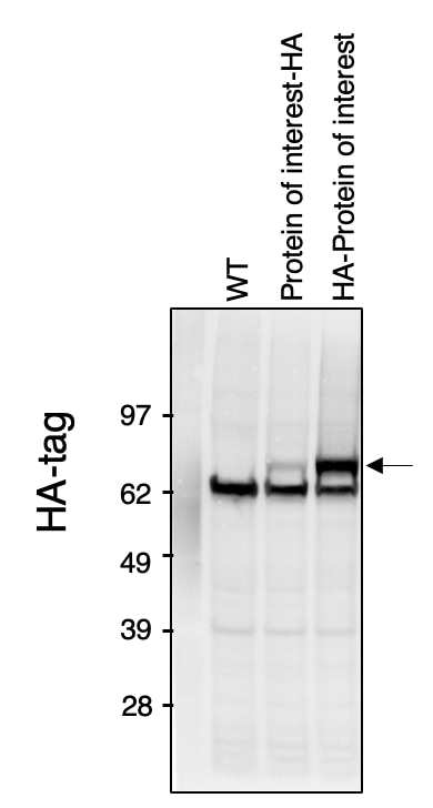
 with Transfected HEK-293 cells lysate 1280 ug.")
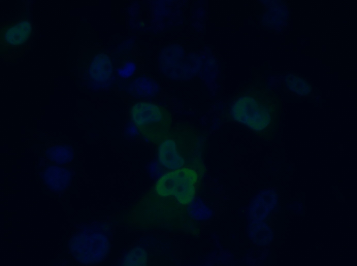
 fixed Transfected HEK-293 cells using HA tag antibody (51064-2-AP) at dilution of 1:200 and CoraLite®488-Conjugated Goat Anti-Rabbit IgG(H+L) (SA00013-2).")
 fixed Transfected HEK-293 cells using HA tag antibody (51064-2-AP) at dilution of 1:200 and CoraLite®594-Conjugated AffiniPure Goat Anti-Rabbit IgG(H+L).")
 and CoraLite®488-Conjugated Goat Anti-Rabbit IgG(H+L) (SA00013-2), and 0.25 ug Isotype Control. Cells were fixed with 4% PFA and permeabilized with Flow Cytometry Perm Buffer (PF00011-C).")
"HA tag Antibodies" Comparison
View side-by-side comparison of HA tag antibodies from other vendors to find the one that best suits your research needs.
Applications testées
| Résultats positifs en WB | Protéine recombinante, cellules HEK-293 transfectées |
| Résultats positifs en IP | cellules HEK-293 transfectées, |
| Résultats positifs en IF/ICC | cellules HEK-293 transfectées, |
| Résultats positifs en FC (Intra) | cellules HEK-293 transfectées, |
Dilution recommandée
| Application | Dilution |
|---|---|
| Western Blot (WB) | WB : 1:5000-1:10000 |
| Immunoprécipitation (IP) | IP : 0.5-4.0 ug for 1.0-3.0 mg of total protein lysate |
| Immunofluorescence (IF)/ICC | IF/ICC : 1:50-1:500 |
| Flow Cytometry (FC) (INTRA) | FC (INTRA) : 0.25 ug per 10^6 cells in a 100 µl suspension |
| It is recommended that this reagent should be titrated in each testing system to obtain optimal results. | |
| Sample-dependent, check data in validation data gallery | |
Applications publiées
| WB | See 778 publications below |
| IF | See 69 publications below |
| IP | See 222 publications below |
| ELISA | See 1 publications below |
| CoIP | See 85 publications below |
| ChIP | See 10 publications below |
Informations sur le produit
51064-2-AP cible HA tag dans les applications de WB, IF/ICC, FC (Intra), IP, CoIP, ChIP, ELISA et montre une réactivité avec des échantillons Protéine recombinante
| Réactivité | Protéine recombinante |
| Réactivité citée | Humain, porc, singe, souris |
| Hôte / Isotype | Lapin / IgG |
| Clonalité | Polyclonal |
| Type | Anticorps |
| Immunogène | Peptide |
| Nom complet | HA tag |
| Masse moléculaire calculée | 1 kDa |
| Symbole du gène | |
| Identification du gène (NCBI) | |
| Conjugaison | Non conjugué |
| Forme | Liquide |
| Méthode de purification | Purification par affinité contre l'antigène |
| Tampon de stockage | PBS with 0.02% sodium azide and 50% glycerol |
| Conditions de stockage | Stocker à -20°C. Stable pendant un an après l'expédition. L'aliquotage n'est pas nécessaire pour le stockage à -20oC Les 20ul contiennent 0,1% de BSA. |
Informations générales
Protein tags are protein or peptide sequences located either on the C- or N- terminal of the target protein, which facilitates one or several of the following characteristics: solubility, detection, purification, localization and expression. The HA tag is corresponds to amino acid residues YPYDVPDYA of human influenza virus hemagglutinin(HA). Many recombinant proteins have been engineered to express the HA tag, which does not appear to interfere with the bioactivity or the biodistribution of the recombinant protein. This tag facilitates the detection, isolation, and purification of the proteins. The HA tag is useful in western blotting and immunohistochemical localization of expressed fusion proteins when examined with antibodies raised specifically against the HA-tag.
Protocole
| Product Specific Protocols | |
|---|---|
| WB protocol for HA tag antibody 51064-2-AP | Download protocol |
| IF protocol for HA tag antibody 51064-2-AP | Download protocol |
| IP protocol for HA tag antibody 51064-2-AP | Download protocol |
| Standard Protocols | |
|---|---|
| Click here to view our Standard Protocols |
Publications
| Species | Application | Title |
|---|---|---|
Signal Transduct Target Ther Circulating tumor cells shielded with extracellular vesicle-derived CD45 evade T cell attack to enable metastasis | ||
Signal Transduct Target Ther FBXW7β loss-of-function enhances FASN-mediated lipogenesis and promotes colorectal cancer growth | ||
Gastroenterology PTEN deficiency facilitates exosome secretion and metastasis in cholangiocarcinoma by impairing TFEB-mediated lysosome biogenesis |
Avis
The reviews below have been submitted by verified Proteintech customers who received an incentive for providing their feedback.
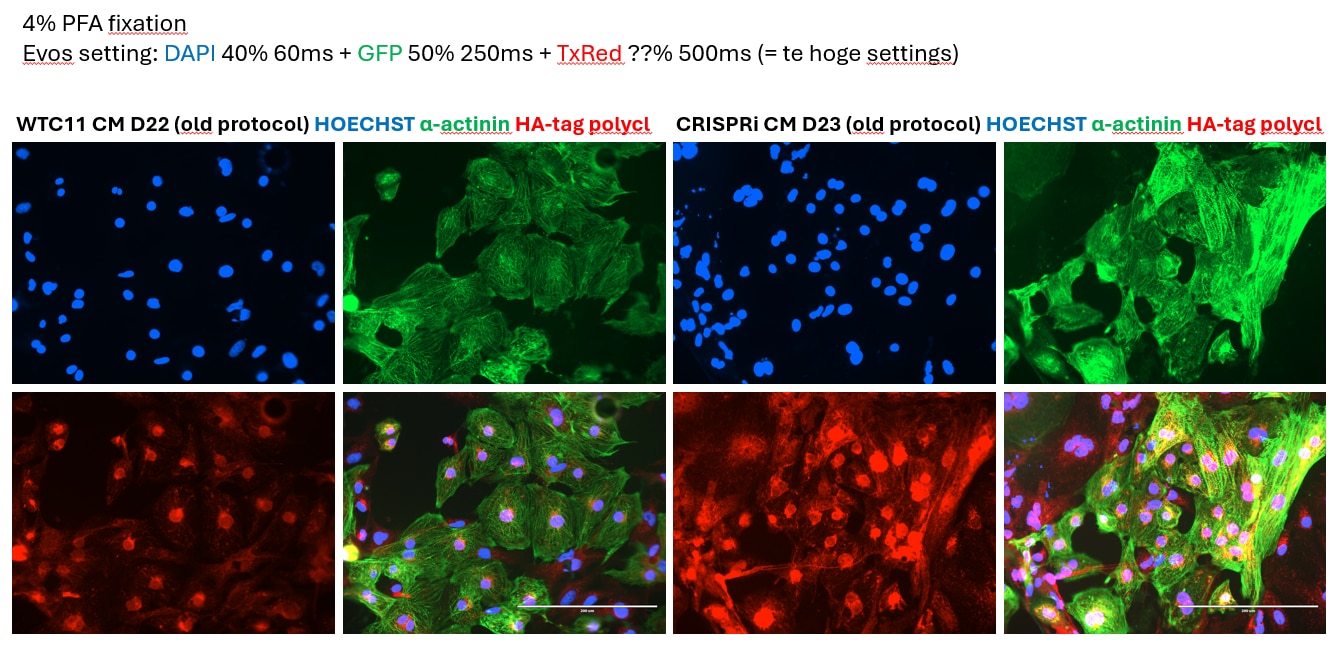
FH Kiara (Verified Customer) (07-28-2025) | This primary antibody did give a signal for HA-tagged dCas9-KRAB in our CRISPRi iPSC-CMs, but however it also gave quite some background in our WTC11 negative control line.
 |
FH Elizabeth (Verified Customer) (07-24-2025) | Our lab mainly purchase Proteintech antibodies. They function well; minimal background "noise". We will continue to purchase these antibodies
|
FH Tiffany (Verified Customer) (05-27-2025) | Very good quality. We dilute this HA antibody in 3% BSA solution and use it as primary antibody for western blot. This mixture can be reused for up to 3 months.
|
FH Tiffany (Verified Customer) (01-06-2025) | The quality is as good as our previous purchase.
|
FH PK (Verified Customer) (07-05-2024) | eXCELLENT
 |
FH S (Verified Customer) (05-16-2024) | Excellent
 |
FH manohar (Verified Customer) (07-10-2023) | Used Nitrocellulose membrane with 5% milk as blocking for 1 hr, 1% milk used for antibody dilution and kept for overnight incubation
|
FH X (Verified Customer) (07-11-2022) | Good for both WB and IF in transfected cells
|
FH Victor (Verified Customer) (06-13-2022) | I'm using the antibody for Western blotting tissue culture samples and it works well for my needs for the price. I've been using a 1:2000 dilution which gives me decent bands via Western blot and analyzed using a fluorescent anti-rabbit secondary. The bands could be a little brighter, but this antibody is worth it for the sale price. It's stored in glycerol, so it saves you the trouble of aliquoting and it can be kept at -20 which is nice.
|
FH Nick (Verified Customer) (04-20-2022) | The antibody works well and performs consistently in IP and WB
|
FH Deyong (Verified Customer) (03-11-2022) | Overall it worked good in the HEK293T cells with protein overexpression, but there were multi non specific bands below.
|
FH Stephanie (Verified Customer) (01-28-2022) | Antibody easy to use, right recommended dilution. No background observed and clean bands.
|
FH S (Verified Customer) (11-22-2021) | Overall, the antibody worked well, but there was a strong band in my control (negative) lane.
 |
FH K (Verified Customer) (07-15-2021) | This ab recognized the HA tag-ed proteins, but also got a lot of non-specific bands, dilution used were 1:2000
|
FH Fen (Verified Customer) (03-06-2020) | it works great for both western blot and IP
|
FH Chun (Verified Customer) (12-05-2019) | An excellent antibody
|
FH Nikhil (Verified Customer) (10-16-2019) | HA tagged VprBP protein was expressed in H1299 cells. Blue: DAPI; Green:HA-VprBP
 |
FH Phuong (Verified Customer) (06-19-2019) | HA tag polyclonal antibody works very well. Gives very clean and strong signal.
|

