- Phare
- Validé par KD/KO
Anticorps Monoclonal anti-HDAC2
HDAC2 Monoclonal Antibody for WB, IHC, IF/ICC, FC (Intra), ELISA
Hôte / Isotype
Mouse / IgG2b
Réactivité testée
Humain, rat, souris
Applications
WB, IHC, IF/ICC, FC (Intra), IP, CoIP, ELISA
Conjugaison
Non conjugué
CloneNo.
1A3E4
N° de cat : 67165-1-Ig
Synonymes
Galerie de données de validation
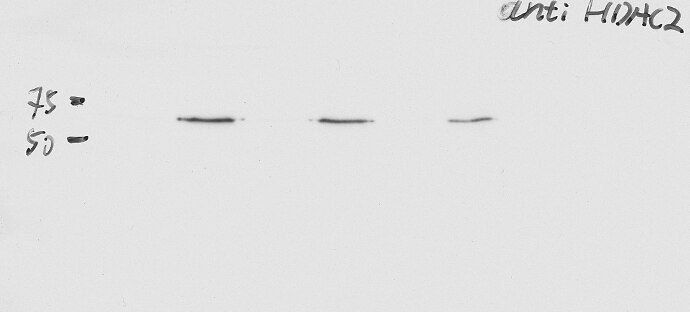
 at dilution of 1:20000 incubated at room temperature for 1.5 hours.")
 at dilution of 1:20000 incubated at room temperature for 1.5 hours.")
 at dilution of 1:1000 (under 10x lens). Heat mediated antigen retrieval with Tris-EDTA buffer (pH 9.0).")
 at dilution of 1:1000 (under 40x lens). Heat mediated antigen retrieval with Tris-EDTA buffer (pH 9.0).")
 fixed HepG2 cells using HDAC2 antibody (67165-1-Ig, Clone: 1A3E4 ) at dilution of 1:800 and CoraLite®488-Conjugated Goat Anti-Mouse IgG(H+L), CL594-Phalloidin (red).")
 and CoraLite®488-Conjugated Goat Anti-Mouse IgG(H+L) at dilution 1:1000 (red), or 0.4 ug Control Antibody. Cells were fixed and permeabilized with Transcription Factor Staining Buffer Kit (PF00011).")
Applications testées
| Résultats positifs en WB | cellules U2OS, cellules 4T1, cellules HEK-293, cellules HeLa, cellules HSC-T6, cellules Jurkat, cellules K-562, cellules MCF-7, cellules NIH/3T3 |
| Résultats positifs en IHC | tissu de cancer du sein humain, il est suggéré de démasquer l'antigène avec un tampon de TE buffer pH 9.0; (*) À défaut, 'le démasquage de l'antigène peut être 'effectué avec un tampon citrate pH 6,0. |
| Résultats positifs en IF/ICC | cellules HepG2, |
| Résultats positifs en FC (Intra) | cellules HepG2 |
| Résultats positifs en cytométrie | cellules HepG2 |
Dilution recommandée
| Application | Dilution |
|---|---|
| Western Blot (WB) | WB : 1:5000-1:50000 |
| Immunohistochimie (IHC) | IHC : 1:500-1:2000 |
| Immunofluorescence (IF)/ICC | IF/ICC : 1:400-1:1600 |
| Flow Cytometry (FC) (INTRA) | FC (INTRA) : 0.40 ug per 10^6 cells in a 100 µl suspension |
| Flow Cytometry (FC) | FC : 0.40 ug per 10^6 cells in a 100 µl suspension |
| It is recommended that this reagent should be titrated in each testing system to obtain optimal results. | |
| Sample-dependent, check data in validation data gallery | |
Applications publiées
| KD/KO | See 2 publications below |
| WB | See 4 publications below |
| IF | See 1 publications below |
| IP | See 1 publications below |
| CoIP | See 2 publications below |
Informations sur le produit
67165-1-Ig cible HDAC2 dans les applications de WB, IHC, IF/ICC, FC (Intra), IP, CoIP, ELISA et montre une réactivité avec des échantillons Humain, rat, souris
| Réactivité | Humain, rat, souris |
| Réactivité citée | rat, Humain |
| Hôte / Isotype | Mouse / IgG2b |
| Clonalité | Monoclonal |
| Type | Anticorps |
| Immunogène | HDAC2 Protéine recombinante Ag21288 |
| Nom complet | histone deacetylase 2 |
| Masse moléculaire calculée | 458 aa, 52 kDa; 488 aa,55 kDa |
| Poids moléculaire observé | 55 kDa |
| Numéro d’acquisition GenBank | BC031055 |
| Symbole du gène | HDAC2 |
| Identification du gène (NCBI) | 3066 |
| Conjugaison | Non conjugué |
| Forme | Liquide |
| Méthode de purification | Purification par protéine A |
| Tampon de stockage | PBS with 0.02% sodium azide and 50% glycerol |
| Conditions de stockage | Stocker à -20°C. Stable pendant un an après l'expédition. L'aliquotage n'est pas nécessaire pour le stockage à -20oC Les 20ul contiennent 0,1% de BSA. |
Informations générales
Histone deacetylases(HDAC) are a class of enzymes that remove the acetyl groups from the lysine residues leading to the formation of a condensed and transcriptionally silenced chromatin.Histone deacetylases act via the formation of large multiprotein complexes, and are responsible for the deacetylation of lysine residues at the N-terminal regions of core histones (H2A, H2B, H3 and H4). At least 4 classes of HDAC were identified. As a class I HDAC, HDAC2 was primarily found in the nucleus. HDAC2 forms transcriptional repressor complexes by associating with many different proteins, including YY1, a mammalian zinc-finger transcription factor. Thus, it plays an important role in transcriptional regulation, cell cycle progression and developmental events. This antibody is raised against residues near the C terminus of human HDAC2.
Protocole
| Product Specific Protocols | |
|---|---|
| WB protocol for HDAC2 antibody 67165-1-Ig | Download protocol |
| IHC protocol for HDAC2 antibody 67165-1-Ig | Download protocol |
| IF protocol for HDAC2 antibody 67165-1-Ig | Download protocol |
| FC protocol for HDAC2 antibody 67165-1-Ig | Download protocol |
| Standard Protocols | |
|---|---|
| Click here to view our Standard Protocols |
Publications
| Species | Application | Title |
|---|---|---|
iScience Modification of lysine-260 2-hydroxyisobutyrylation destabilizes ALDH1A1 expression to regulate bladder cancer progression
| ||
Molecules Anti-Colorectal Cancer Activity of Solasonin from Solanum nigrum L. via Histone Deacetylases-Mediated p53 Acetylation Pathway | ||
Toxicol Appl Pharmacol Advanced oxidation protein products upregulate ABCB1 expression and activity via HDAC2-Foxo3α-mediated signaling in vitro and in vivo.
| ||
Theranostics CRISPR screening reveals ZNF217 as a vulnerability in high-risk B-cell acute lymphoblastic leukemia |
Avis
The reviews below have been submitted by verified Proteintech customers who received an incentive for providing their feedback.
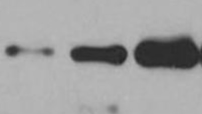
FH Xinda (Verified Customer) (02-24-2025) | human iPSC derived NPC cell lysates were used as input. Co-immunoprecipitation was performed against a protein of interest. On the graph, one can see that the beads coupled with control antibody did not pull out HDAC2, whereas the protein of interest pulled out HDAC2. The groups are not labeled (because our results are not published), but in triplicate.
 |
FH Xiaoyu (Verified Customer) (06-28-2023) | Good for wb
 |

