- Phare
- Validé par KD/KO
Anticorps Monoclonal anti-HIF-1 alpha
HIF-1 alpha Monoclonal Antibody for WB, IHC, ELISA
Hôte / Isotype
Mouse / IgG1
Réactivité testée
Humain
Applications
WB, IHC, IF, IP, CoIP, ChIP, ELISA
Conjugaison
Non conjugué
CloneNo.
1H3C12
N° de cat : 66730-1-Ig
Synonymes
Galerie de données de validation
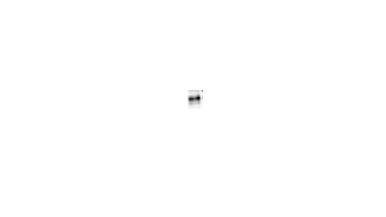
 at dilution of 1:10000 incubated at room temperature for 1.5 hours. The membrane was stripped and re-blotted with YY1 antibody as loading control.")
 at dilution of 1:200 (under 40x lens). Heat mediated antigen retrieval with Tris-EDTA buffer (pH 9.0).")
 at dilution of 1:200 (under 40x lens). Heat mediated antigen retrieval with Tris-EDTA buffer (pH 9.0).")
 at dilution of 1:200 (under 10x lens). Heat mediated antigen retrieval with Tris-EDTA buffer (pH 9.0).")
 at dilution of 1:200 (under 10x lens). Heat mediated antigen retrieval with Tris-EDTA buffer (pH 9.0).")
Applications testées
| Résultats positifs en WB | cellules HeLa, cellules HeLa traitées au chlorure de cobalt, cellules HepG2 traitées au chlorure de cobalt |
| Résultats positifs en IHC | tissu de cancer du poumon humain, tissu de tumeur ovarienne humain il est suggéré de démasquer l'antigène avec un tampon de TE buffer pH 9.0; (*) À défaut, 'le démasquage de l'antigène peut être 'effectué avec un tampon citrate pH 6,0. |
Dilution recommandée
| Application | Dilution |
|---|---|
| Western Blot (WB) | WB : 1:2000-1:10000 |
| Immunohistochimie (IHC) | IHC : 1:50-1:500 |
| It is recommended that this reagent should be titrated in each testing system to obtain optimal results. | |
| Sample-dependent, check data in validation data gallery | |
Applications publiées
| KD/KO | See 3 publications below |
| WB | See 61 publications below |
| IHC | See 15 publications below |
| IF | See 17 publications below |
| IP | See 3 publications below |
| CoIP | See 1 publications below |
| ChIP | See 1 publications below |
Informations sur le produit
66730-1-Ig cible HIF-1 alpha dans les applications de WB, IHC, IF, IP, CoIP, ChIP, ELISA et montre une réactivité avec des échantillons Humain
| Réactivité | Humain |
| Réactivité citée | Humain |
| Hôte / Isotype | Mouse / IgG1 |
| Clonalité | Monoclonal |
| Type | Anticorps |
| Immunogène | HIF-1 alpha Protéine recombinante Ag15198 |
| Nom complet | hypoxia inducible factor 1, alpha subunit (basic helix-loop-helix transcription factor) |
| Masse moléculaire calculée | 826 aa, 93 kDa |
| Poids moléculaire observé | 120 kDa |
| Numéro d’acquisition GenBank | BC012527 |
| Symbole du gène | HIF1A |
| Identification du gène (NCBI) | 3091 |
| Conjugaison | Non conjugué |
| Forme | Liquide |
| Méthode de purification | Purification par protéine G |
| Tampon de stockage | PBS with 0.02% sodium azide and 50% glycerol |
| Conditions de stockage | Stocker à -20°C. Stable pendant un an après l'expédition. L'aliquotage n'est pas nécessaire pour le stockage à -20oC Les 20ul contiennent 0,1% de BSA. |
Informations générales
HIF1a, the major regulator of the cellular responses to hypoxia, consists of an oxygen-sensitive subunit, HIF1 alpha (HIF1A), and an oxygen-insensitive subunit, HIF1 beta (arylhydrocarbon receptor nuclear transporter [ARNT]). Under normal oxygen conditions, HIF1a is continuously produced and destroyed, in a process involving hydroxylation, interaction with von Hippel-Lindau (VHL) protein, polyubiquitylation and subsequent proteasomal degradation. Under hypoxic conditions, hydroxylation is impaired and HIF1a is stabilized. HIF1a localizes in cytoplasm in normoxia, but it can translocate into nuclear in response to hypoxia. The calculated molecular weight of HIF1a is 93 kDa, but the modified protein HIF1a is about 110-120kDa (PMID: 11698256, .PMID: 7539918). .
Protocole
| Product Specific Protocols | |
|---|---|
| WB protocol for HIF-1 alpha antibody 66730-1-Ig | Download protocol |
| IHC protocol for HIF-1 alpha antibody 66730-1-Ig | Download protocol |
| Standard Protocols | |
|---|---|
| Click here to view our Standard Protocols |
Publications
| Species | Application | Title |
|---|---|---|
Adv Sci (Weinh) Transgelin Promotes Glioblastoma Stem Cell Hypoxic Responses and Maintenance Through p53 Acetylation | ||
Theranostics ITGB2-mediated metabolic switch in CAFs promotes OSCC proliferation by oxidation of NADH in mitochondrial oxidative phosphorylation system. | ||
Phytomedicine mTOR/HIF-1α pathway-mediated glucose reprogramming and macrophage polarization by Sini decoction plus ginseng soup in ALF | ||
Oxid Med Cell Longev Chitosan Oligosaccharides Alleviate H2O2-stimulated Granulosa Cell Damage via HIF-1α Signaling Pathway. |
Avis
The reviews below have been submitted by verified Proteintech customers who received an incentive for providing their feedback.
FH S (Verified Customer) (05-16-2024) | Excellent
 |
FH balawant (Verified Customer) (12-20-2022) | I have used this antibody for western blot and it is working great
|
FH Diane (Verified Customer) (02-02-2021) | incubated over night 4 degrees C. 1:2500 HRP conjugated secondary. Opti-4CN substrate kit for detection. Also tried ICC on cytospins so ran out of trial size before fully optimized but has potential to provide the information that I am needing.
 |

