- Phare
- Validé par KD/KO
Anticorps Polyclonal de lapin anti-Histone H3
Histone H3 Polyclonal Antibody for WB, IHC, IF/ICC, FC (Intra), IP, ELISA
Hôte / Isotype
Lapin / IgG
Réactivité testée
Humain, rat, souris et plus (6)
Applications
WB, IHC, IF/ICC, FC (Intra), IP, CoIP, ChIP, ELISA
Conjugaison
Non conjugué
N° de cat : 17168-1-AP
Synonymes
 at dilution of 1:8000 incubated at room temperature for 1.5 hours.")
 at dilution of 1:1000 incubated at room temperature for 1.5 hours.")
 at dilution of 1:300 incubated at room temperature for 1.5 hours.")
 at dilution of 1:300 incubated at room temperature for 1.5 hours.")
 at dilution of 1:300 incubated at room temperature for 1.5 hours.")
 with MCF-7 cells lysate 2120 ug.")
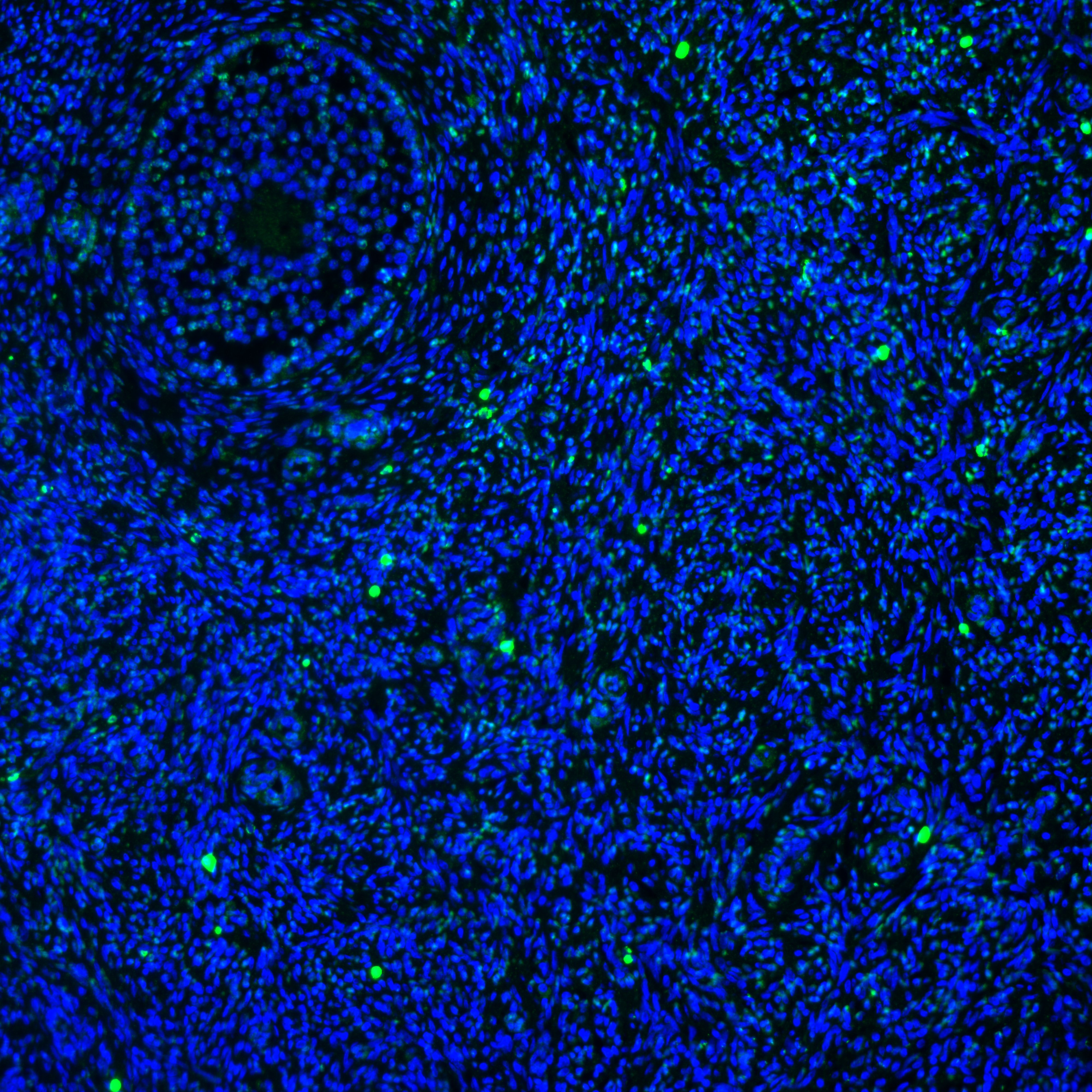
 at dilution of 1:200 (under 40x lens). Heat mediated antigen retrieval with Tris-EDTA buffer (pH 9.0).")
 at dilution of 1:200 (under 10x lens). Heat mediated antigen retrieval with Tris-EDTA buffer (pH 9.0).")
 at dilution of 1:200 (under 40x lens).")
 at dilution of 1:200 (under 10x lens).")
 at dilution of 1:2000 (under 10x lens). Heat mediated antigen retrieval with Tris-EDTA buffer (pH 9.0).")
 at dilution of 1:2000 (under 40x lens). Heat mediated antigen retrieval with Tris-EDTA buffer (pH 9.0).")
 fixed HeLa cells using Histone-H3 antibody (17168-1-AP) at dilution of 1:200 and CoraLite®488-Conjugated AffiniPure Goat Anti-Rabbit IgG(H+L).")
 fixed HeLa cells using Histone H3 antibody (17168-1-AP) at dilution of 1:1200 and CoraLite®488-Conjugated AffiniPure Goat Anti-Rabbit IgG(H+L), Beta Actin antibody (66009-1-Ig, Clone: 2D4H5, red).")
 fixed HeLa cells using Histone-H3 antibody (17168-1-AP) at dilution of 1:200 and CoraLite®488-Conjugated AffiniPure Goat Anti-Rabbit IgG(H+L), CL594-Phalloidin (red).")
 at dilution of 1:50 and Rhodamine-Goat anti-Rabbit IgG.")
 and CoraLite®488-Conjugated Goat Anti-Rabbit IgG(H+L) (SA00013-2)(red), or 0.25 ug rabbit IgG isotype control (blue). Cells were fixed with 4% PFA and permeabilized with Flow Cytometry Perm Buffer (PF00011-C).")
"Histone H3 Antibodies" Comparison
View side-by-side comparison of Histone H3 antibodies from other vendors to find the one that best suits your research needs.
Applications testées
| Résultats positifs en WB | cellules HEK-293, cellules A549, cellules HeLa, cellules HepG, cellules MCF-7, cellules NIH/3T3, tissu cérébral de souris, tissu de muscle squelettique de souris, tissu hépatique de souris, tissu rénal de rat, tissu rénal de souris |
| Résultats positifs en IP | cellules MCF-7, |
| Résultats positifs en IHC | tissu de cancer de l'œsophage humain, tissu de cancer de la peau humain, tissu de cancer du sein humain il est suggéré de démasquer l'antigène avec un tampon de TE buffer pH 9.0; (*) À défaut, 'le démasquage de l'antigène peut être 'effectué avec un tampon citrate pH 6,0. |
| Résultats positifs en IF/ICC | cellules HeLa, |
| Résultats positifs en FC (Intra) | cellules HeLa, |
Dilution recommandée
| Application | Dilution |
|---|---|
| Western Blot (WB) | WB : 1:2000-1:16000 |
| Immunoprécipitation (IP) | IP : 0.5-4.0 ug for 1.0-3.0 mg of total protein lysate |
| Immunohistochimie (IHC) | IHC : 1:50-1:500 |
| Immunofluorescence (IF)/ICC | IF/ICC : 1:600-1:2400 |
| Flow Cytometry (FC) (INTRA) | FC (INTRA) : 0.25 ug per 10^6 cells in a 100 µl suspension |
| It is recommended that this reagent should be titrated in each testing system to obtain optimal results. | |
| Sample-dependent, check data in validation data gallery | |
Applications publiées
| KD/KO | See 2 publications below |
| WB | See 1005 publications below |
| IHC | See 3 publications below |
| IF | See 10 publications below |
| IP | See 1 publications below |
| CoIP | See 2 publications below |
| ChIP | See 9 publications below |
Informations sur le produit
17168-1-AP cible Histone H3 dans les applications de WB, IHC, IF/ICC, FC (Intra), IP, CoIP, ChIP, ELISA et montre une réactivité avec des échantillons Humain, rat, souris
| Réactivité | Humain, rat, souris |
| Réactivité citée | rat, Chèvre, Humain, porc, poulet, singe, souris, arabidopsis, fish |
| Hôte / Isotype | Lapin / IgG |
| Clonalité | Polyclonal |
| Type | Anticorps |
| Immunogène | Histone H3 Protéine recombinante Ag10644 |
| Nom complet | histone cluster 2, H3a |
| Masse moléculaire calculée | 136 aa, 15 kDa |
| Poids moléculaire observé | 15-17 kDa |
| Numéro d’acquisition GenBank | BC015544 |
| Symbole du gène | Histone H3 |
| Identification du gène (NCBI) | 333932 |
| Conjugaison | Non conjugué |
| Forme | Liquide |
| Méthode de purification | Purification par affinité contre l'antigène |
| Tampon de stockage | PBS with 0.02% sodium azide and 50% glycerol |
| Conditions de stockage | Stocker à -20°C. Stable pendant un an après l'expédition. L'aliquotage n'est pas nécessaire pour le stockage à -20oC Les 20ul contiennent 0,1% de BSA. |
Informations générales
Histone-H3, histone cluster 2, H3a is the core component of nucleosome. Nucleosomes wrap and compact DNA into chromatin, limiting DNA accessibility to the cellular machinery which requires DNA as a template. Histones thereby play a central role in transcription regulation, DNA repair, DNA replication and chromosomal stability. DNA accessibility is regulated via a complex set of post-translational modifications of histones, also called histone code, and nucleosome remodeling. Histone-H3 is expressed during S phase; then expression strongly decreases as cell division slows down during the process of differentiation.
Protocole
| Product Specific Protocols | |
|---|---|
| WB protocol for Histone H3 antibody 17168-1-AP | Download protocol |
| IHC protocol for Histone H3 antibody 17168-1-AP | Download protocol |
| IF protocol for Histone H3 antibody 17168-1-AP | Download protocol |
| IP protocol for Histone H3 antibody 17168-1-AP | Download protocol |
| Standard Protocols | |
|---|---|
| Click here to view our Standard Protocols |
Publications
| Species | Application | Title |
|---|---|---|
Signal Transduct Target Ther TRAF3 activates STING-mediated suppression of EV-A71 and target of viral evasion | ||
Mol Cancer Cell surface CD55 traffics to the nucleus leading to cisplatin resistance and stemness by inducing PRC2 and H3K27 trimethylation on chromatin in ovarian cancer | ||
Cell Targeting Epigenetic Crosstalk as a Therapeutic Strategy for EZH2-Aberrant Solid Tumors. |
Avis
The reviews below have been submitted by verified Proteintech customers who received an incentive for providing their feedback.
FH Felipe (Verified Customer) (07-24-2025) | Histone H3 antibody is a reliable marker for detecting proliferating cells. After optimizing the dilution, I achieved good results in identifying proliferative cells in bovine ovarian tissue using an immunofluorescence assay.
 |
FH Christin (Verified Customer) (06-14-2025) | Antibody worked very vell for western blot using iPSC-derived macrophages and microglia
|
FH kis (Verified Customer) (02-21-2025) | This antibody gave u satisfactory results in our cell line for Western blot (WB) analysis.
|
FH Parijat (Verified Customer) (08-19-2024) | Worked on blot.
|
FH Wissem (Verified Customer) (06-27-2024) | Antibody works very well at detecting Histone H3 in extracts from U2OS, HCT116, HeLa, RPE1 and HEK293 cells - used at 1:10,000 to 1:15,000 incubated overnight at 4C
|
FH Roy (Verified Customer) (06-12-2024) | This antibody is great at detecting Histone H3 by WB (1/1000- 1h incubation at RT) with beautiful enrichment at the purified chromatin fraction.
|
FH Mi (Verified Customer) (06-25-2023) | It works very well in nuclear extracts from human brown adipocyte cells.
|
FH Veda (Verified Customer) (04-29-2022) | IF staining test in cryo sections of skin. It does not seem to work. Maybe it only stains the nuclear membrane.
|
FH Pradeep (Verified Customer) (09-19-2020) | Good antibody for Western blot. Selvaraju, V., Thirunavukkarasu, M., Joshi, M. et al. Deletion of newly described pro-survival molecule Pellino-1 increases oxidative stress, downregulates cIAP2/NF-κB cell survival pathway, reduces angiogenic response, and thereby aggravates tissue function in mouse ischemic models. Basic Res Cardiol 115, 45 (2020). https://doi.org/10.1007/s00395-020-0804-4
|
FH Tanusree (Verified Customer) (12-03-2019) | The antibody works great in Western blotting analysis.
|
FH Ruiting (Verified Customer) (11-11-2019) | I use the Proteintech antibodies almost every week, the antibodies never failed to me. Very gald to use their products with a great price.Ruiting
|
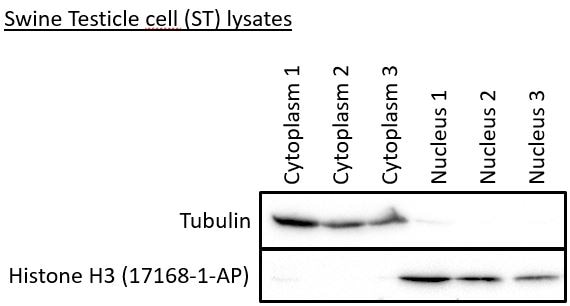
FH Robert (Verified Customer) (07-18-2019) | The Histon H3 antibody gives a signal in the three nuclear samples, but not in the cytoplasmic samples.
 |

