- Phare
- Validé par KD/KO
Anticorps Polyclonal de lapin anti-IBA1
IBA1 Polyclonal Antibody for IHC, IF-P, FC (Intra), ELISA
Hôte / Isotype
Lapin / IgG
Réactivité testée
Humain, rat, souris et plus (4)
Applications
IHC, IF-P, FC (Intra), ELISA
Conjugaison
Non conjugué
N° de cat : 10904-1-AP
Synonymes
Galerie de données de validation
 with THP-1 cells lysate 2400 ug.")
 at dilution of 1:4000 (under 40x lens). Heat mediated antigen retrieval with Tris-EDTA buffer (pH 9.0).")
 at dilution of 1:4000 (under 10x lens). Heat mediated antigen retrieval with Tris-EDTA buffer (pH 9.0).")
 at dilution of 1:1000 (under 10x lens). Heat mediated antigen retrieval with Tris-EDTA buffer (pH 9.0).")
 at dilution of 1:1000 (under 40x lens). Heat mediated antigen retrieval with Tris-EDTA buffer (pH 9.0).")
 at dilution of 1:1000 (under 10x lens). Heat mediated antigen retrieval with Tris-EDTA buffer (pH 9.0).")
 at dilution of 1:1000 (under 40x lens). Heat mediated antigen retrieval with Tris-EDTA buffer (pH 9.0).")
 at dilution of 1:800 (under 10x lens). Heat mediated antigen retrieval with Tris-EDTA buffer (pH 9.0).")
 at dilution of 1:800 (under 40x lens). Heat mediated antigen retrieval with Tris-EDTA buffer (pH 9.0).")
 at dilution of 1:1000 (under 10x lens). Heat mediated antigen retrieval with Tris-EDTA buffer (pH 9.0).")
 at dilution of 1:1000 (under 40x lens). Heat mediated antigen retrieval with Tris-EDTA buffer (pH 9.0).")
 fixed mouse brain tissue using IBA1 antibody (10904-1-AP) at dilution of 1:200 and CoraLite®488-Conjugated AffiniPure Goat Anti-Rabbit IgG(H+L).")
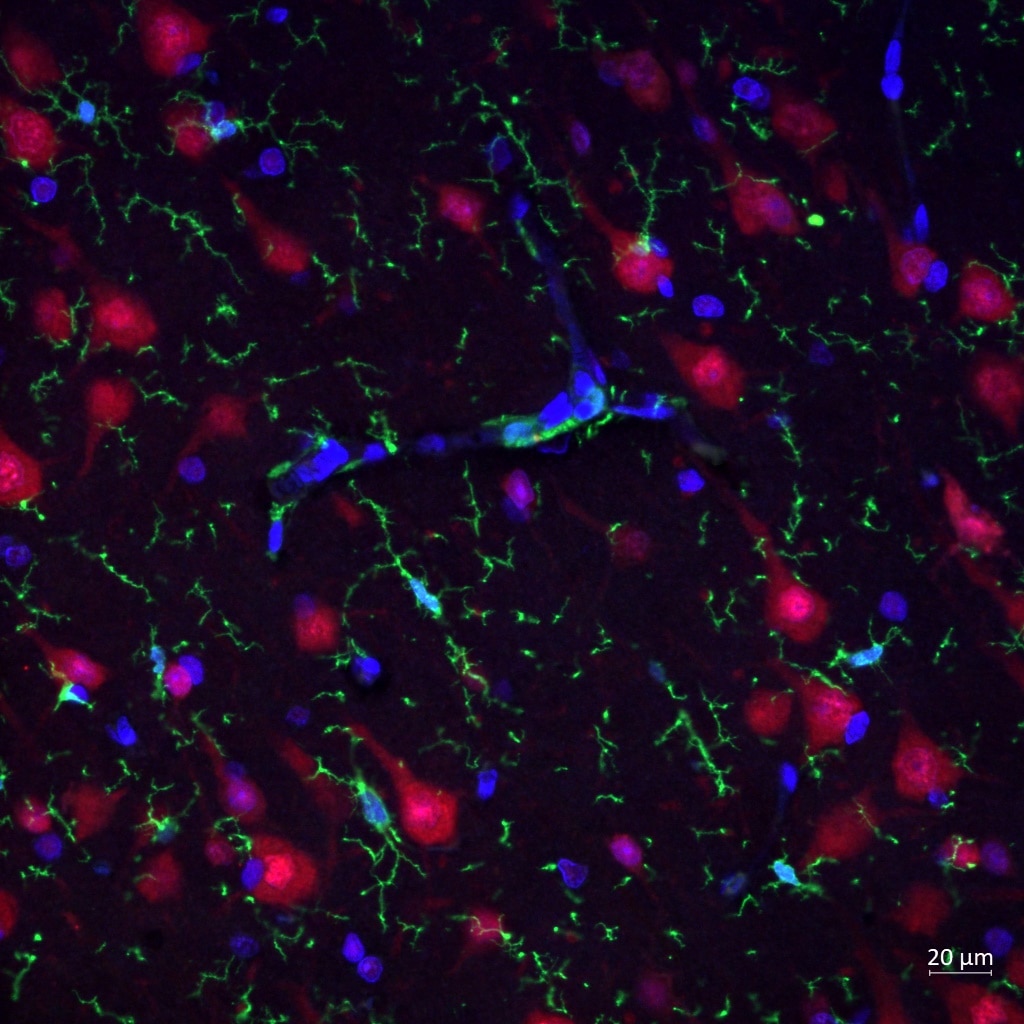
 fixed rat brain tissue using IBA1 antibody (10904-1-AP) at dilution of 1:200 and CoraLite®488-Conjugated AffiniPure Goat Anti-Rabbit IgG(H+L), MAP2 antibody (17490-1-AP, orange), GFAP antibody (60190-1-Ig, Clone: 4B2E10, Magenta).")
 fixed frozen OCT-embedded mouse brain tissue using IBA1 antibody (10904-1-AP) at dilution of 1:200 and CoraLite®488-Conjugated Goat Anti-Rabbit IgG(H+L) (SA00013-2).")
 and CoraLite®488-Conjugated AffiniPure Goat Anti-Rabbit IgG(H+L) at dilution 1:1000 (red), or 0.4 ug Control Antibody. Cells were fixed with 4% PFA and permeabilized with Flow Cytometry Perm Buffer (PF00011-C).")
Applications testées
| Résultats positifs en IP | cellules THP-1, |
| Résultats positifs en IHC | tissu cérébral de rat, tissu cérébral humain, tissu d'amygdalite humain, tissu hépatique humain il est suggéré de démasquer l'antigène avec un tampon de TE buffer pH 9.0; (*) À défaut, 'le démasquage de l'antigène peut être 'effectué avec un tampon citrate pH 6,0. |
| Résultats positifs en IF-P | tissu cérébral de souris, tissu cérébral de rat |
| Résultats positifs en IF-Fro | tissu cérébral de souris, |
| Résultats positifs en FC (Intra) | cellules THP-1 |
Dilution recommandée
| Application | Dilution |
|---|---|
| Immunoprécipitation (IP) | IP : 0.5-4.0 ug for 1.0-3.0 mg of total protein lysate |
| Immunohistochimie (IHC) | IHC : 1:2000-1:8000 |
| Immunofluorescence (IF)-P | IF-P : 1:50-1:500 |
| Immunofluorescence (IF)-FRO | IF-FRO : 1:50-1:500 |
| Flow Cytometry (FC) (INTRA) | FC (INTRA) : 0.40 ug per 10^6 cells in a 100 µl suspension |
| It is recommended that this reagent should be titrated in each testing system to obtain optimal results. | |
| Sample-dependent, check data in validation data gallery | |
Applications publiées
| KD/KO | See 5 publications below |
| WB | See 1 publications below |
| IHC | See 109 publications below |
| IF | See 279 publications below |
Informations sur le produit
10904-1-AP cible IBA1 dans les applications de IHC, IF-P, FC (Intra), ELISA et montre une réactivité avec des échantillons Humain, rat, souris
| Réactivité | Humain, rat, souris |
| Réactivité citée | rat, Humain, poisson-zèbre, porc, singe, souris, Hamster |
| Hôte / Isotype | Lapin / IgG |
| Clonalité | Polyclonal |
| Type | Anticorps |
| Immunogène | IBA1 Protéine recombinante Ag1363 |
| Nom complet | allograft inflammatory factor 1 |
| Masse moléculaire calculée | 17 kDa |
| Numéro d’acquisition GenBank | BC009474 |
| Symbole du gène | IBA1 |
| Identification du gène (NCBI) | 199 |
| Conjugaison | Non conjugué |
| Forme | Liquide |
| Méthode de purification | Purification par affinité contre l'antigène |
| Tampon de stockage | PBS with 0.02% sodium azide and 50% glycerol |
| Conditions de stockage | Stocker à -20°C. Stable pendant un an après l'expédition. L'aliquotage n'est pas nécessaire pour le stockage à -20oC Les 20ul contiennent 0,1% de BSA. |
Informations générales
What is the molecular weight of IBA1?
The molecular weight of IBA1 is 16.7 kD.
What is IBA1?
Ionized calcium-binding adaptor molecule 1 (IBA1), also known as Allograft inflammatory factor-1 (AIF-1), is an inflammation-responsive scaffold protein expressed and secreted from macrophages. Microglia response factor (MRF-1) and daintain are also similar, and likely identical, proteins (PMIDs: 29749461, 9630473, 23792284).
What is the function of IBA1?
IBA1 is necessary for macrophage survival, and it is also a key molecule in proinflammatory activity (PMID: 29749461).
Where is IBA1 localized?
IBA1 is a cytoplasmic protein, often expressed in immunocytes, macrophages, and microglia. It can be used as a marker for normal (not 'dark') microglia in brain tissue, as IBA1 is expressed by all microglial cell subpopulations (PMIDs: 29749461, 11943136, 26847266, 9630473).
Is IBA1 upregulated in active immunophages?
Yes; its expression is associated with inflammatory activity (PMID: 29749461).
How do IBA1-positive microglia differ from microglia that express less or no IBA1?
'Dark' microglia is a recently described phenotype associated with Alzheimer's disease pathology and chronic stress; these dark microglia express decreased IBA1 (often punctiform), and show distinct ultrastructural differences from 'normal' microglia as well as condensed and electron-dense cytoplasm and nucleoplasm. Normal microglia generally display strong, diffuse expression of IBA1. IBA1-positive microglial processes in normal conditions also have a strong tendency to exclusively contact synaptic elements, such as axon terminals, dendritic spines, and synaptic clefts (PMIDs: 29992181, 26847266).
How is IBA1 related to Alzheimer's disease and pain?
Exacerbated immunoactivity of IBA1, particularly in proximity to amyloid plaques, is prominently featured in AD pathology. IBA1, along with other microglial markers, are also associated with pain, and robust microglial reactions often follow spinal cord injury (PMIDs: 29992181, 23792284).
Protocole
| Product Specific Protocols | |
|---|---|
| IHC protocol for IBA1 antibody 10904-1-AP | Download protocol |
| IF protocol for IBA1 antibody 10904-1-AP | Download protocol |
| IP protocol for IBA1 antibody 10904-1-AP | Download protocol |
| Standard Protocols | |
|---|---|
| Click here to view our Standard Protocols |
Publications
| Species | Application | Title |
|---|---|---|
Int J Surg Stereotactically intracerebral transplantation of neural stem cells for ischemic stroke attenuated inflammatory responses and promoted neurogenesis: an experimental study with monkeys | ||
Adv Sci (Weinh) OTUD5 Protects Dopaminergic Neurons by Promoting the Degradation of α-Synuclein in Parkinson's Disease Model | ||
Int J Surg Bioinformatics analysis identifies CSF1R as an essential gene mediating Neuropathic pain - Experimental research. | ||
Cell Death Differ Redox regulation of TRIM28 facilitates neuronal ferroptosis by promoting SUMOylation and inhibiting OPTN-selective autophagic degradation of ACSL4 |
Avis
The reviews below have been submitted by verified Proteintech customers who received an incentive for providing their feedback.
FH Reyes (Verified Customer) (04-05-2024) | Iba1 (in green) marks clearly my microglia in human brain FFPE cortex.
 |
FH Alexandru (Verified Customer) (11-13-2023) | Very pleased with the antibody! I had a claer signal in all my stains.
|
FH NX (Verified Customer) (02-27-2023) | A clean antibody to detect endogenous Iba1 from mouse brain lysate.
|

