Anticorps Monoclonal anti-human IBA1
human IBA1 Monoclonal Antibody for IHC, ELISA
Hôte / Isotype
Mouse / IgG2a
Réactivité testée
Humain
Applications
WB, IHC, IF, ELISA
Conjugaison
Non conjugué
CloneNo.
1C6A10
N° de cat : 66827-1-Ig
Synonymes
Galerie de données de validation
 at dilution of 1:4000 (under 40x lens. Heat mediated antigen retrieval with Tris-EDTA buffer (pH 9.0).")
 at dilution of 1:4000 (under 10x lens. Heat mediated antigen retrieval with Tris-EDTA buffer (pH 9.0).")
 at dilution of 1:1000 (under 40x lens. Heat mediated antigen retrieval with Tris-EDTA buffer (pH 9.0).")
 at dilution of 1:1000 (under 10x lens. Heat mediated antigen retrieval with Tris-EDTA buffer (pH 9.0).")
 at dilution of 1:1000 (under 10x lens. Heat mediated antigen retrieval with Tris-EDTA buffer (pH 9.0).")
 at dilution of 1:1000 (under 40x lens. Heat mediated antigen retrieval with Tris-EDTA buffer (pH 9.0).")
 at dilution of 1:1000 (under 40x lens. Heat mediated antigen retrieval with Tris-EDTA buffer (pH 9.0).")
Applications testées
| Résultats positifs en IHC | tissu cérébral humain, tissu d'amygdalite humain, tissu de cancer du foie humain, tissu de gliome humain il est suggéré de démasquer l'antigène avec un tampon de TE buffer pH 9.0; (*) À défaut, 'le démasquage de l'antigène peut être 'effectué avec un tampon citrate pH 6,0. |
Dilution recommandée
| Application | Dilution |
|---|---|
| Immunohistochimie (IHC) | IHC : 1:1000-1:4000 |
| It is recommended that this reagent should be titrated in each testing system to obtain optimal results. | |
| Sample-dependent, check data in validation data gallery | |
Applications publiées
| WB | See 8 publications below |
| IHC | See 6 publications below |
| IF | See 40 publications below |
Informations sur le produit
66827-1-Ig cible human IBA1 dans les applications de WB, IHC, IF, ELISA et montre une réactivité avec des échantillons Humain
| Réactivité | Humain |
| Réactivité citée | Humain |
| Hôte / Isotype | Mouse / IgG2a |
| Clonalité | Monoclonal |
| Type | Anticorps |
| Immunogène | human IBA1 Protéine recombinante Ag28236 |
| Nom complet | allograft inflammatory factor 1 |
| Masse moléculaire calculée | 17 kDa |
| Numéro d’acquisition GenBank | BC009474 |
| Symbole du gène | IBA1 |
| Identification du gène (NCBI) | 199 |
| Conjugaison | Non conjugué |
| Forme | Liquide |
| Méthode de purification | Purification par protéine A |
| Tampon de stockage | PBS with 0.02% sodium azide and 50% glycerol |
| Conditions de stockage | Stocker à -20°C. Stable pendant un an après l'expédition. L'aliquotage n'est pas nécessaire pour le stockage à -20oC Les 20ul contiennent 0,1% de BSA. |
Informations générales
IBA1 is a 143 amino acid cytoplasmic, inflammation response scaffold protein. It is constitutively expressed in monocytes and macrophages and is known to be involved in macrophage activation. It is a marker of activated macrophage. Expression of IBA1 is up-regulated in activated microglia following facial nerve axotomy, ischemia, and several brain diseases, thereby implicating it in the activated phenotypes of microglia.
Protocole
| Product Specific Protocols | |
|---|---|
| IHC protocol for human IBA1 antibody 66827-1-Ig | Download protocol |
| Standard Protocols | |
|---|---|
| Click here to view our Standard Protocols |
Publications
| Species | Application | Title |
|---|---|---|
Nat Commun Fatal iatrogenic cerebral β-amyloid-related arteritis in a woman treated with lecanemab for Alzheimer's disease | ||
Cell Death Dis ChemR23 activation attenuates cognitive impairment in chronic cerebral hypoperfusion by inhibiting NLRP3 inflammasome-induced neuronal pyroptosis | ||
Phytomedicine Withaferin A protects against epilepsy by promoting LCN2-mediated astrocyte polarization to stopping neuronal ferroptosis | ||
Oxid Med Cell Longev Senkyunolide I Protects against Sepsis-Associated Encephalopathy by Attenuating Sleep Deprivation in a Murine Model of Cecal Ligation and Puncture. | ||
Neural Regen Res Mitochondrial transplantation confers protection against the effects of ischemic stroke by repressing microglial pyroptosis and promoting neurogenesis | ||
J Neuroinflammation Upregulation of TRPC5 in hippocampal excitatory synapses improves memory impairment associated with neuroinflammation in microglia knockout IL-10 mice. |
Avis
The reviews below have been submitted by verified Proteintech customers who received an incentive for providing their feedback.
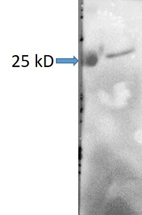
FH Nuc (Verified Customer) (10-24-2023) | There is a clear band at 25 kD which is not the right MW.
 |

