Anticorps Recombinant de lapin anti-IBA1
IBA1 Recombinant Antibody for WB, IHC, IHC-Autostainer, IF-P, FC (Intra), ELISA
Hôte / Isotype
Lapin / IgG
Réactivité testée
Humain, rat, souris
Applications
WB, IHC, IHC-Autostainer, IF-P, FC (Intra), ELISA
Conjugaison
Non conjugué
CloneNo.
4D5
N° de cat : 81728-1-RR
Synonymes
Galerie de données de validation
 at dilution of 1:10000 incubated at room temperature for 1.5 hours.")
 at dilution of 1:10000 incubated at room temperature for 1.5 hours.")
 at dilution of 1:200 (under 40x lens). Heat mediated antigen retrieval with Citrate buffer (pH 6.0). Staining was performed on the Epredia 480S Autostainer.")
 at dilution of 1:200 (under 10x lens). Heat mediated antigen retrieval with Citrate buffer (pH 6.0). Staining was performed on the Epredia 480S Autostainer.")
 at dilution of 1:1000 (under 40x lens). Heat mediated antigen retrieval with Tris-EDTA buffer (pH 9.0).")
 at dilution of 1:1000 (under 10x lens). Heat mediated antigen retrieval with Tris-EDTA buffer (pH 9.0).")
 at dilution of 1:1000 (under 10x lens). Heat mediated antigen retrieval with Tris-EDTA buffer (pH 9.0).")
 at dilution of 1:1000 (under 40x lens). Heat mediated antigen retrieval with Tris-EDTA buffer (pH 9.0).")
 at dilution of 1:1000 (under 10x lens). Heat mediated antigen retrieval with Tris-EDTA buffer (pH 9.0).")
 at dilution of 1:1000 (under 40x lens). Heat mediated antigen retrieval with Tris-EDTA buffer (pH 9.0).")
 at dilution of 1:400 (under 10x lens). Heat mediated antigen retrieval with Tris-EDTA buffer (pH 9.0).")
 at dilution of 1:400 (under 40x lens). Heat mediated antigen retrieval with Tris-EDTA buffer (pH 9.0).")
 fixed rat brain tissue using IBA1 antibody (81728-1-RR, Clone: 4D5 ) at dilution of 1:500 and CoraLite®488-Conjugated AffiniPure Goat Anti-Rabbit IgG(H+L).")
 fixed rat brain tissue using IBA1 antibody (81728-1-RR, Clone: 4D5 ) at dilution of 1:500 and CoraLite®488-Conjugated AffiniPure Goat Anti-Rabbit IgG(H+L).")
 fixed paraffin-embedded rat brain tissue using IBA1 antibody (81728-1-RR, Clone: 4D5 ) at dilution of 1:200 and Multi-rAb CoraLite ® Plus 750-Goat Anti-Rabbit Recombinant Secondary Antibody (H+L) (RGAR006). Heat mediated antigen retrieval with Tris-EDTA buffer (pH 9.0).")
 fixed paraffin-embedded rat brain tissue using IBA1 antibody (81728-1-RR, Clone: 4D5 ) at dilution of 1:250 and CoraLite®488-Conjugated Goat Anti-Rabbit IgG(H+L) (SA00013-2). Heat mediated antigen retrieval with Tris-EDTA buffer (pH 9.0).")
 and CoraLite®488-Conjugated AffiniPure Goat Anti-Rabbit IgG(H+L) (SA00013-2)(red), or 0.4 ug Isotype Control (blue). Cells were fixed with 4% PFA and permeabilized with Flow Cytometry Perm Buffer (PF00011-C).")
Applications testées
| Résultats positifs en WB | cellules THP-1, cellules HL-60, cellules U-937 |
| Résultats positifs en IHC-Autostainer | tissu amygdalien humain, |
| Résultats positifs en IHC | tissu cérébral de rat, tissu amygdalien humain, tissu cérébral de souris, tissu hépatique de souris, tissu hépatique humain il est suggéré de démasquer l'antigène avec un tampon de TE buffer pH 9.0; (*) À défaut, 'le démasquage de l'antigène peut être 'effectué avec un tampon citrate pH 6,0. |
| Résultats positifs en IF-P | tissu cérébral de rat, |
| Résultats positifs en FC (Intra) | cellules THP-1 |
Dilution recommandée
| Application | Dilution |
|---|---|
| Western Blot (WB) | WB : 1:5000-1:50000 |
| Immunohistochimie (IHC)-AUTOSTAINER | IHC-AUTOSTAINER : 1:50-1:500 |
| Immunohistochimie (IHC) | IHC : 1:500-1:2000 |
| Immunofluorescence (IF)-P | IF-P : 1:250-1:1000 |
| Flow Cytometry (FC) (INTRA) | FC (INTRA) : 0.40 ug per 10^6 cells in a 100 µl suspension |
| It is recommended that this reagent should be titrated in each testing system to obtain optimal results. | |
| Sample-dependent, check data in validation data gallery | |
Applications publiées
| IHC | See 2 publications below |
| IF | See 5 publications below |
Informations sur le produit
81728-1-RR cible IBA1 dans les applications de WB, IHC, IHC-Autostainer, IF-P, FC (Intra), ELISA et montre une réactivité avec des échantillons Humain, rat, souris
| Réactivité | Humain, rat, souris |
| Réactivité citée | rat, souris |
| Hôte / Isotype | Lapin / IgG |
| Clonalité | Recombinant |
| Type | Anticorps |
| Immunogène | IBA1 Protéine recombinante Ag1363 |
| Nom complet | allograft inflammatory factor 1 |
| Masse moléculaire calculée | 17 kDa |
| Poids moléculaire observé | 17 kDa |
| Numéro d’acquisition GenBank | BC009474 |
| Symbole du gène | IBA1 |
| Identification du gène (NCBI) | 199 |
| Conjugaison | Non conjugué |
| Forme | Liquide |
| Méthode de purification | Purification par protéine A |
| Tampon de stockage | PBS with 0.02% sodium azide and 50% glycerol |
| Conditions de stockage | Stocker à -20°C. Stable pendant un an après l'expédition. L'aliquotage n'est pas nécessaire pour le stockage à -20oC Les 20ul contiennent 0,1% de BSA. |
Informations générales
IBA1 is a 143 amino acid cytoplasmic, inflammation response scaffold protein. It is constitutively expressed in monocytes and macrophages and is known to be involved in macrophage activation. It is a marker of activated macrophage. Expression of IBA1 is up-regulated in activated microglia following facial nerve axotomy, ischemia, and several brain diseases, thereby implicating it in the activated phenotypes of microglia.
Protocole
| Product Specific Protocols | |
|---|---|
| WB protocol for IBA1 antibody 81728-1-RR | Download protocol |
| IHC protocol for IBA1 antibody 81728-1-RR | Download protocol |
| IF protocol for IBA1 antibody 81728-1-RR | Download protocol |
| Standard Protocols | |
|---|---|
| Click here to view our Standard Protocols |
Publications
| Species | Application | Title |
|---|---|---|
Exp Cell Res Targeting of ubiquitination and degradation of KLF15 by E3 ubiquitin ligase KBTBD7 regulates LPS-induced septic brain injury in microglia | ||
Antioxidants (Basel) Withania somnifera (Ashwagandha) Improves Spatial Memory, Anxiety and Depressive-like Behavior in the 5xFAD Mouse Model of Alzheimer's Disease | ||
CNS Neurosci Ther A Novel Compound Ligusticum Cycloprolactam Alleviates Neuroinflammation After Ischemic Stroke via the FPR1/NLRP3 Signaling Axis | ||
Brain Behav The Activation of PKM2 Induces Pyroptosis in Hippocampal Neurons via the NLRP3/Caspase-1/GSDMD Pathway in Neonatal Rats With Hypoxic-Ischemic Brain Injury | ||
Adv Mater Biomimetic Nanomotors for Deep Ischemia Penetration and Ferroptosis Inhibition in Neuroprotective Therapy of Ischemic Stroke | ||
Int Immunopharmacol Macrophage migration inhibitory factor in the mouse hippocampus promotes neuroinflammation and cognitive dysfunction following anesthesia and surgery |
Avis
The reviews below have been submitted by verified Proteintech customers who received an incentive for providing their feedback.
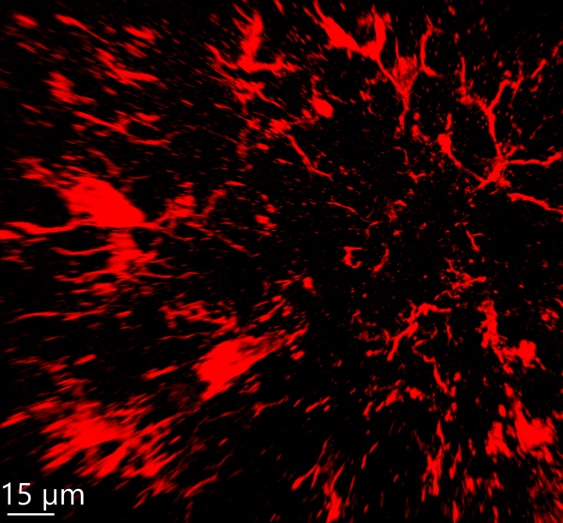
FH Kateryna (Verified Customer) (01-23-2025) | IBA1 was used as a microglial-specific marker to visualize microglia in mouse brain tissue, specifically the cortex. The antibody worked consistently well on the fixed tissue: 40-μm thick sections, 3% normal goat serum blocking, and overnight incubation with the antibody (IBA1). In the image, red represents IBA1 visualized by an Alexa 633-labeled anti-rabbit fluorescent secondary antibody.
 |

