- Phare
- Validé par KD/KO
Anticorps Monoclonal anti-ICAM-1/CD54
ICAM-1/CD54 Monoclonal Antibody for IF/ICC, IF-P
Hôte / Isotype
Mouse / IgG2b
Réactivité testée
Humain
Applications
IF/ICC, IF-P
Conjugaison
CoraLite®594 Fluorescent Dye
CloneNo.
2F9A8
N° de cat : CL594-60299
Synonymes
Galerie de données de validation
Applications testées
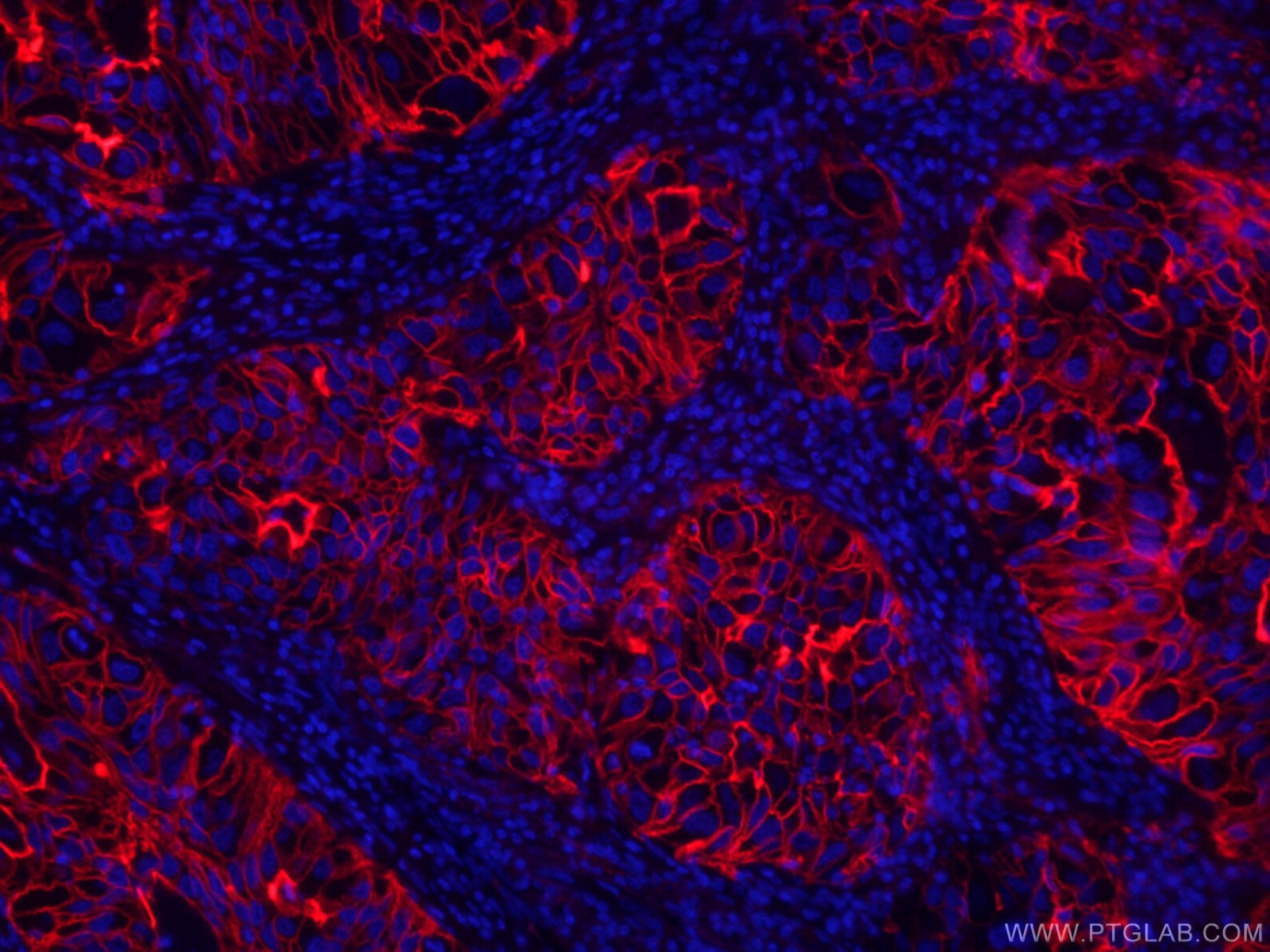
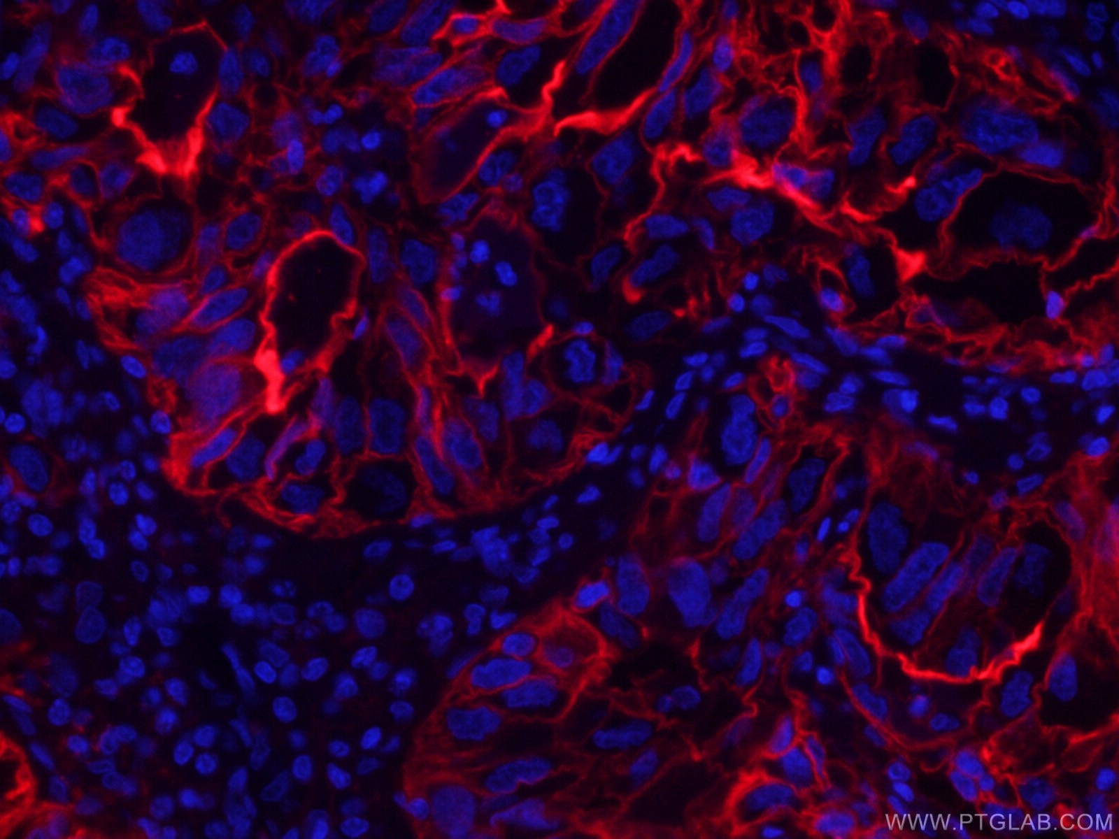
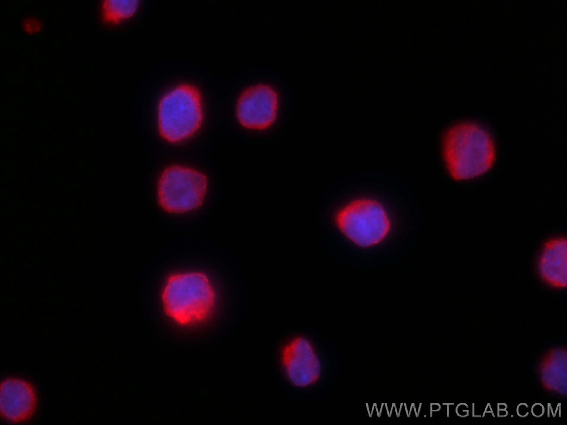
| Résultats positifs en IF-P | tissu de cancer du poumon humain, cellules Raji |
| Résultats positifs en IF/ICC | cellules Raji, |
Dilution recommandée
| Application | Dilution |
|---|---|
| Immunofluorescence (IF)-P | IF-P : 1:50-1:500 |
| Immunofluorescence (IF)/ICC | IF/ICC : 1:50-1:500 |
| It is recommended that this reagent should be titrated in each testing system to obtain optimal results. | |
| Sample-dependent, check data in validation data gallery | |
Informations sur le produit
CL594-60299 cible ICAM-1/CD54 dans les applications de IF/ICC, IF-P et montre une réactivité avec des échantillons Humain
| Réactivité | Humain |
| Hôte / Isotype | Mouse / IgG2b |
| Clonalité | Monoclonal |
| Type | Anticorps |
| Immunogène | ICAM-1/CD54 Protéine recombinante Ag8309 |
| Nom complet | intercellular adhesion molecule 1 |
| Masse moléculaire calculée | 90 kDa |
| Poids moléculaire observé | 85-95 kDa |
| Numéro d’acquisition GenBank | BC015969 |
| Symbole du gène | ICAM-1 |
| Identification du gène (NCBI) | 3383 |
| Conjugaison | CoraLite®594 Fluorescent Dye |
| Excitation/Emission maxima wavelengths | 588 nm / 604 nm |
| Forme | Liquide |
| Méthode de purification | Purification par protéine A |
| Tampon de stockage | PBS with 50% glycerol, 0.05% Proclin300, 0.5% BSA |
| Conditions de stockage | Stocker à -20 °C. Éviter toute exposition à la lumière. Stable pendant un an après l'expédition. L'aliquotage n'est pas nécessaire pour le stockage à -20oC Les 20ul contiennent 0,1% de BSA. |
Informations générales
Where is ICAM-1 expressed?
Intercellular Adhesion Molecule 1 (ICAM-1), also known as Cluster of Differentiation 54 (CD54) is a transmembrane glycoprotein constitutively expressed at low levels in endothelial cells, pericytes and on some lymphocytes and monocytes1. It is located at the cytoplasmic membrane, with a large extracellular region of mainly hydrophobic amino acids joined to a small transmembrane region and a cytoplasmic tail. It has a molecular weight of 75 to 115 kDa depending on the level of glycosylation.
What is the function of ICAM-1?
ICAM-1 is important in both innate and adaptive immune responses as an adhesion molecule. Although it is constitutively expressed, in the presence of pro-inflammatory cytokines such as TNFα the endothelial cells are activated and upregulate expression of ICAM-12. In blood vessels lined with endothelial cells, leukocytes that are rolling over the surface are able to bind to ICAM-1 and transmigrate through the endothelial barrier and into the tissue. The initial binding of the leukocytes to ICAM-1 causes a Ca2+ release that initiates endothelial cell contraction and weakening of the intercellular tight junctions3, 4. This protein can be used as an indicator of endothelial activation and of vascular inflammation.
What is the role of ICAM-1 in disease?
Beyond the role in the immune response, ICAM-1 has also been identified as the target of attachment for the human rhinovirus, the cause of the common cold. Binding of the virus to ICAM-1 causes the viral capsid to uncoat and leads to release of the genetic material5.
Hubbard, A. K. & Rothlein, R. Intercellular adhesion molecule-1 (ICAM-1) expression and cell signaling cascades. Free Radic. Biol. Med. 28, 1379-86 (2000).
Long, E. O. ICAM-1: getting a grip on leukocyte adhesion. J. Immunol. 186, 5021-3 (2011).
Lawson, C. & Wolf, S. ICAM-1 signaling in endothelial cells. (2009).
Lyck, R. & Enzmann, G. The physiological roles of ICAM-1 and ICAM-2 in neutrophil migration into tissues. Curr. Opin. Hematol. 22, 53-59 (2015).
Xing, L., Casasnovas, J. M. & Cheng, R. H. Structural analysis of human rhinovirus complexed with ICAM-1 reveals the dynamics of receptor-mediated virus uncoating. J. Virol. 77, 6101-7 (2003).
Protocole
| Product Specific Protocols | |
|---|---|
| IF protocol for CL594 ICAM-1/CD54 antibody CL594-60299 | Download protocol |
| Standard Protocols | |
|---|---|
| Click here to view our Standard Protocols |


