- Phare
- Validé par KD/KO
Anticorps Polyclonal de lapin anti-MAEA
MAEA Polyclonal Antibody for WB, IHC, ELISA
Hôte / Isotype
Lapin / IgG
Réactivité testée
Humain et plus (1)
Applications
WB, IHC, IP, ELISA
Conjugaison
Non conjugué
N° de cat : 28363-1-AP
Synonymes
Galerie de données de validation
 at dilution of 1:2000 incubated at room temperature for 1.5 hours.")
 at dilution of 1:200 (under 10x lens). Heat mediated antigen retrieval with Tris-EDTA buffer (pH 9.0).")
 at dilution of 1:200 (under 40x lens). Heat mediated antigen retrieval with Tris-EDTA buffer (pH 9.0).")
 at dilution of 1:200 (under 10x lens). Heat mediated antigen retrieval with Tris-EDTA buffer (pH 9.0).")
 at dilution of 1:200 (under 40x lens). Heat mediated antigen retrieval with Tris-EDTA buffer (pH 9.0).")
Applications testées
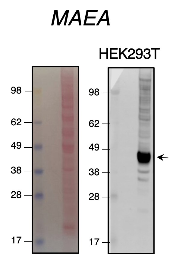
| Résultats positifs en WB | cellules HepG2, cellules Jurkat |
| Résultats positifs en IHC | tissu de cancer du foie humain, tissu de cancer du sein humain il est suggéré de démasquer l'antigène avec un tampon de TE buffer pH 9.0; (*) À défaut, 'le démasquage de l'antigène peut être 'effectué avec un tampon citrate pH 6,0. |
Dilution recommandée
| Application | Dilution |
|---|---|
| Western Blot (WB) | WB : 1:1000-1:4000 |
| Immunohistochimie (IHC) | IHC : 1:50-1:500 |
| It is recommended that this reagent should be titrated in each testing system to obtain optimal results. | |
| Sample-dependent, check data in validation data gallery | |
Applications publiées
| KD/KO | See 2 publications below |
| WB | See 7 publications below |
| IHC | See 2 publications below |
| IP | See 1 publications below |
Informations sur le produit
28363-1-AP cible MAEA dans les applications de WB, IHC, IP, ELISA et montre une réactivité avec des échantillons Humain
| Réactivité | Humain |
| Réactivité citée | Humain, souris |
| Hôte / Isotype | Lapin / IgG |
| Clonalité | Polyclonal |
| Type | Anticorps |
| Immunogène | MAEA Protéine recombinante Ag27150 |
| Nom complet | macrophage erythroblast attacher |
| Masse moléculaire calculée | 45 kDa |
| Poids moléculaire observé | 45 kDa |
| Numéro d’acquisition GenBank | BC001225 |
| Symbole du gène | MAEA |
| Identification du gène (NCBI) | 10296 |
| Conjugaison | Non conjugué |
| Forme | Liquide |
| Méthode de purification | Purification par affinité contre l'antigène |
| Tampon de stockage | PBS with 0.02% sodium azide and 50% glycerol |
| Conditions de stockage | Stocker à -20°C. Stable pendant un an après l'expédition. L'aliquotage n'est pas nécessaire pour le stockage à -20oC Les 20ul contiennent 0,1% de BSA. |
Informations générales
MAEA(macrophage erythroblast attacher) is a core component of the CTLH E3 ubiquitin-protein ligase complex. MAEA is required for normal cell proliferation and plays a role in erythroblast enucleation during erythrocyte maturation and in the development of mature macrophages. N-terminus MAEA and full-length MAEA are mostly expressed in nuclear and can be detected at low level in the cytoplasm of some cells, while C-terminus MAEA can be detected in the cytoplasm and nucleoli within the nucleus. MAEA mediates the attachment of erythroid cell to mature macrophages which can inhibit erythroid cell apoptosis. The expression of MAEA may be associated with the type 2 diabetes mellitus.(PubMed: 28955747, 24143168, 29911972, 29911972, 30674470, 9763581).
Protocole
| Product Specific Protocols | |
|---|---|
| WB protocol for MAEA antibody 28363-1-AP | Download protocol |
| IHC protocol for MAEA antibody 28363-1-AP | Download protocol |
| Standard Protocols | |
|---|---|
| Click here to view our Standard Protocols |
Publications
| Species | Application | Title |
|---|---|---|
Cancers (Basel) Oncopeptide MBOP Encoded by LINC01234 Promotes Colorectal Cancer through MAPK Signaling Pathway. | ||
Oncogene E3 ligase MAEA-mediated ubiquitination and degradation of PHD3 promotes glioblastoma progression
| ||
Chembiochem Identifying E3 ligase substrates with quantitative degradation proteomics
| ||
J Transl Med LAD1 promotes malignant progression by diminishing ubiquitin-dependent degradation of vimentin in gastric cancer | ||
Mol Cell mTORC1-CTLH E3 ligase regulates the degradation of HMG-CoA synthase 1 through the Pro/N-degron pathway | ||
Int J Biol Sci The E3 ubiquitin ligase MAEA promotes macrophage phagocytosis and inhibits gastrointestinal cancer progression by mediating PARP1 ubiquitination and degradation |
Avis
The reviews below have been submitted by verified Proteintech customers who received an incentive for providing their feedback.
FH S (Verified Customer) (12-31-2021) | Quite a good quality antibody. KO validated.
 |
FH H (Verified Customer) (07-09-2020) | This is very good antibody. MAEA band is very thick and clear.
 |

