Anticorps Recombinant de lapin anti-MAP2
MAP2 Recombinant Antibody for IF-P
Hôte / Isotype
Lapin / IgG
Réactivité testée
Humain, rat, souris
Applications
IF-P
Conjugaison
CoraLite®594 Fluorescent Dye
CloneNo.
241653E9
N° de cat : CL594-84306-3
Synonymes
Galerie de données de validation
Applications testées
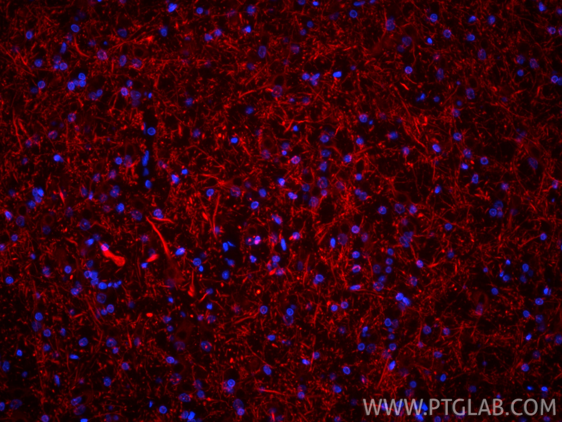
| Résultats positifs en IF-P | tissu cérébral de rat, |
Dilution recommandée
| Application | Dilution |
|---|---|
| Immunofluorescence (IF)-P | IF-P : 1:50-1:500 |
| It is recommended that this reagent should be titrated in each testing system to obtain optimal results. | |
| Sample-dependent, check data in validation data gallery | |
Informations sur le produit
CL594-84306-3 cible MAP2 dans les applications de IF-P et montre une réactivité avec des échantillons Humain, rat, souris
| Réactivité | Humain, rat, souris |
| Hôte / Isotype | Lapin / IgG |
| Clonalité | Recombinant |
| Type | Anticorps |
| Immunogène | MAP2 Protéine recombinante Ag11580 |
| Nom complet | microtubule-associated protein 2 |
| Masse moléculaire calculée | 200 kDa |
| Poids moléculaire observé | 280-300 kDa, 70-85 kDa |
| Numéro d’acquisition GenBank | BC038857 |
| Symbole du gène | MAP2 |
| Identification du gène (NCBI) | 4133 |
| Conjugaison | CoraLite®594 Fluorescent Dye |
| Excitation/Emission maxima wavelengths | 588 nm / 604 nm |
| Forme | Liquide |
| Méthode de purification | Purification par protéine A |
| Tampon de stockage | PBS with 50% glycerol, 0.05% Proclin300, 0.5% BSA |
| Conditions de stockage | Stocker à -20 °C. Éviter toute exposition à la lumière. Stable pendant un an après l'expédition. L'aliquotage n'est pas nécessaire pour le stockage à -20oC Les 20ul contiennent 0,1% de BSA. |
Informations générales
MAP2 (microtubule-associated protein 2) is a cytoskeleton protein abundant in the brain and has an important role in neuronal morphogenesis. Multiple high MW and low MW MAP2 isoforms are expressed within the proximal segment of axons, dendrites, and cell bodies. The expression of MAP2 is regulated in both a tissue- and developmentally-specific manner. The 280 kDa MAP2B is present throughout rat brain development, and the slightly larger MAP2A appears first during the end of the second week of postnatal life. MAP2C, composed of several bands of about 70 kDa, is present during early brain development and largely disappears from the mature brain except for the retina, olfactory bulb, and cerebellum. In addition, some isoforms with lower MW around 50-60 kDa also exist. MAP2 antibodies have been widely used to mark the neuron or dendrite formation.
Protocole
| Product Specific Protocols | |
|---|---|
| IF protocol for CL594 MAP2 antibody CL594-84306-3 | Download protocol |
| Standard Protocols | |
|---|---|
| Click here to view our Standard Protocols |


