Anticorps Monoclonal anti-MFF
MFF Monoclonal Antibody for WB, IHC, IF-P, ELISA
Hôte / Isotype
Mouse / IgG1
Réactivité testée
Humain, porc, rat, souris
Applications
WB, IHC, IF-P, ELISA
Conjugaison
Non conjugué
CloneNo.
1G4B12
N° de cat : 66527-1-Ig
Synonymes
Galerie de données de validation
Applications testées
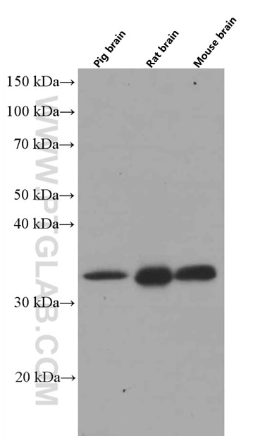
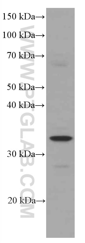
| Résultats positifs en WB | tissu cérébral de porc, cellules HepG2, cellules L02, tissu cérébral de rat, tissu cérébral de souris |
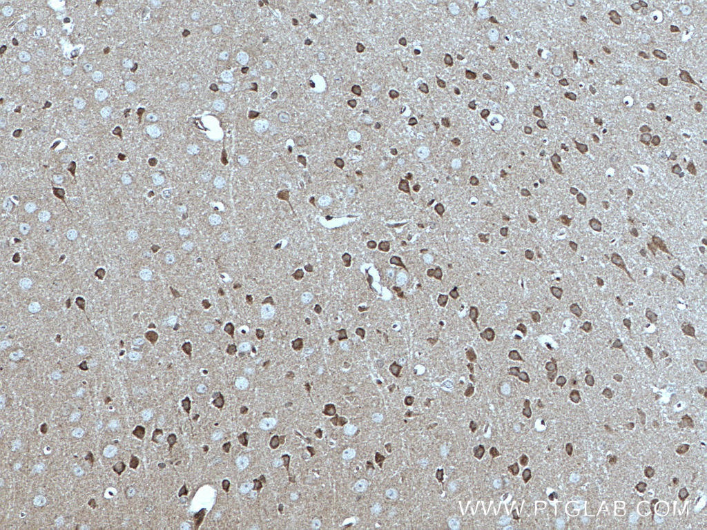
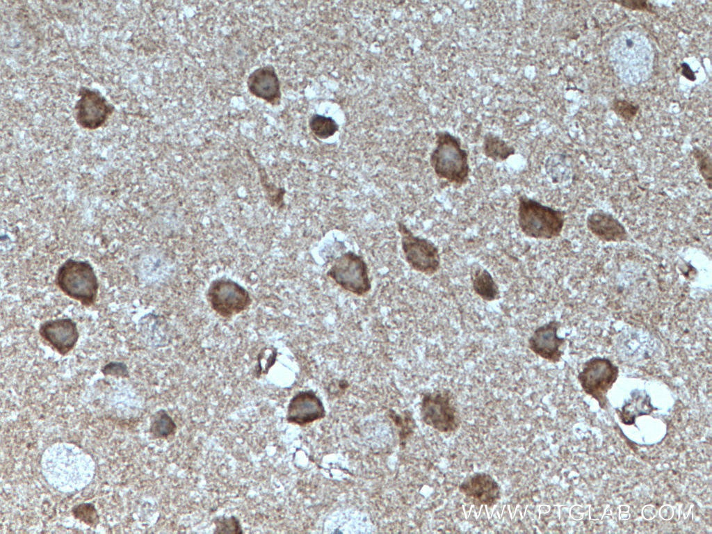
| Résultats positifs en IHC | tissu cérébral de souris, il est suggéré de démasquer l'antigène avec un tampon de TE buffer pH 9.0; (*) À défaut, 'le démasquage de l'antigène peut être 'effectué avec un tampon citrate pH 6,0. |
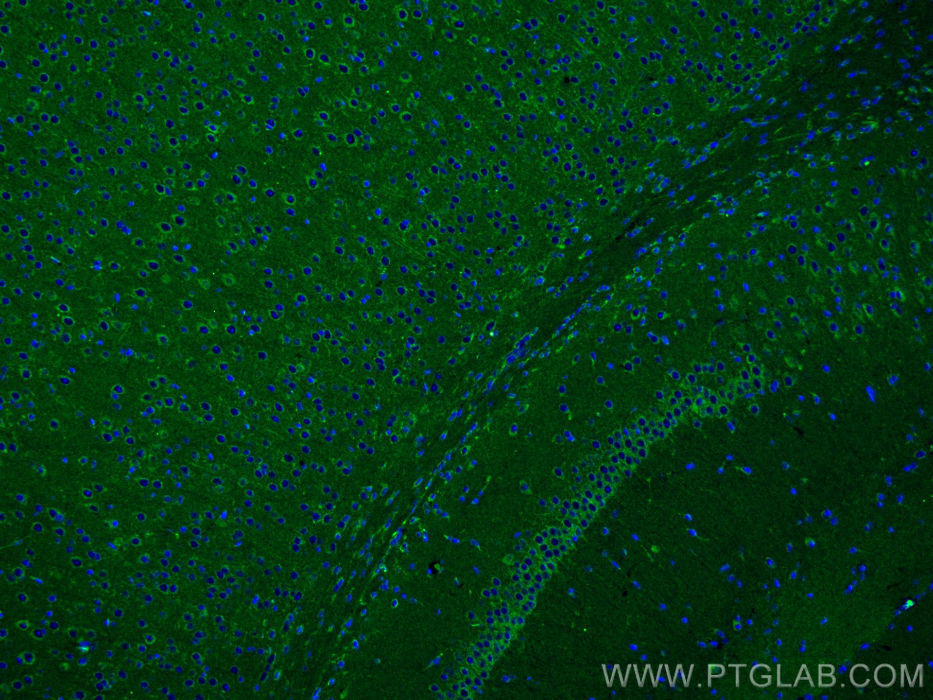
| Résultats positifs en IF-P | tissu cérébral de souris, |
Dilution recommandée
| Application | Dilution |
|---|---|
| Western Blot (WB) | WB : 1:2000-1:10000 |
| Immunohistochimie (IHC) | IHC : 1:250-1:1000 |
| Immunofluorescence (IF)-P | IF-P : 1:200-1:800 |
| It is recommended that this reagent should be titrated in each testing system to obtain optimal results. | |
| Sample-dependent, check data in validation data gallery | |
Applications publiées
| WB | See 6 publications below |
| IHC | See 1 publications below |
| IF | See 2 publications below |
Informations sur le produit
66527-1-Ig cible MFF dans les applications de WB, IHC, IF-P, ELISA et montre une réactivité avec des échantillons Humain, porc, rat, souris
| Réactivité | Humain, porc, rat, souris |
| Réactivité citée | rat, Humain, souris |
| Hôte / Isotype | Mouse / IgG1 |
| Clonalité | Monoclonal |
| Type | Anticorps |
| Immunogène | MFF Protéine recombinante Ag9990 |
| Nom complet | mitochondrial fission factor |
| Masse moléculaire calculée | 38 kDa |
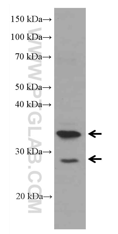
| Poids moléculaire observé | 35-38 kDa |
| Numéro d’acquisition GenBank | BC000797 |
| Symbole du gène | MFF |
| Identification du gène (NCBI) | 56947 |
| Conjugaison | Non conjugué |
| Forme | Liquide |
| Méthode de purification | Purification par protéine G |
| Tampon de stockage | PBS with 0.02% sodium azide and 50% glycerol |
| Conditions de stockage | Stocker à -20°C. Stable pendant un an après l'expédition. L'aliquotage n'est pas nécessaire pour le stockage à -20oC Les 20ul contiennent 0,1% de BSA. |
Informations générales
MFF (mitochondrial fission factor) is a mitochondrial outer membrane protein that is involved in mitochondrial localization of Drp1 and mitochondrial fission. Multiple isoforms of MFF exist due to the alternative splicing. This antibody recognizes the endogenous MFF protein around 26-29 kDa and 35-38 kDa.
Protocole
| Product Specific Protocols | |
|---|---|
| WB protocol for MFF antibody 66527-1-Ig | Download protocol |
| IHC protocol for MFF antibody 66527-1-Ig | Download protocol |
| IF protocol for MFF antibody 66527-1-Ig | Download protocol |
| Standard Protocols | |
|---|---|
| Click here to view our Standard Protocols |
Publications
| Species | Application | Title |
|---|---|---|
Phytother Res Shenlian extract decreases mitochondrial autophagy to regulate mitochondrial function in microvascular to alleviate coronary artery no-reflow | ||
J Adv Res Ceramide-1-phosphate alleviates high-altitude pulmonary edema by stabilizing circadian ARNTL-mediated mitochondrial dynamics | ||
iScience Deficient tRNA posttranscription modification dysregulated the mitochondrial quality controls and apoptosis | ||
Theriogenology OPA1 deficiency induces mitophagy through PINK1/Parkin pathway during bovine oocytes maturation | ||
Respir Res Activated DRP1 promotes mitochondrial fission and induces glycolysis in ATII cells under hyperoxia |
Avis
The reviews below have been submitted by verified Proteintech customers who received an incentive for providing their feedback.
FH JAE HO (Verified Customer) (05-06-2019) | 25 ug of PC3 cell lysates were used for western blot.500 ug of cell lysates and 2.5 ug of antibodies were used for IP.Comparing to "17090-1-AP", it looks less titer but it works well for WB and IP.
|
