- Phare
- Validé par KD/KO
Anticorps Polyclonal de lapin anti-MFN1
MFN1 Polyclonal Antibody for WB, IHC, IF/ICC, IP, ELISA
Hôte / Isotype
Lapin / IgG
Réactivité testée
Humain, rat, souris et plus (7)
Applications
WB, IHC, IF/ICC, IP, ELISA
Conjugaison
Non conjugué
N° de cat : 13798-1-AP
Synonymes
Galerie de données de validation
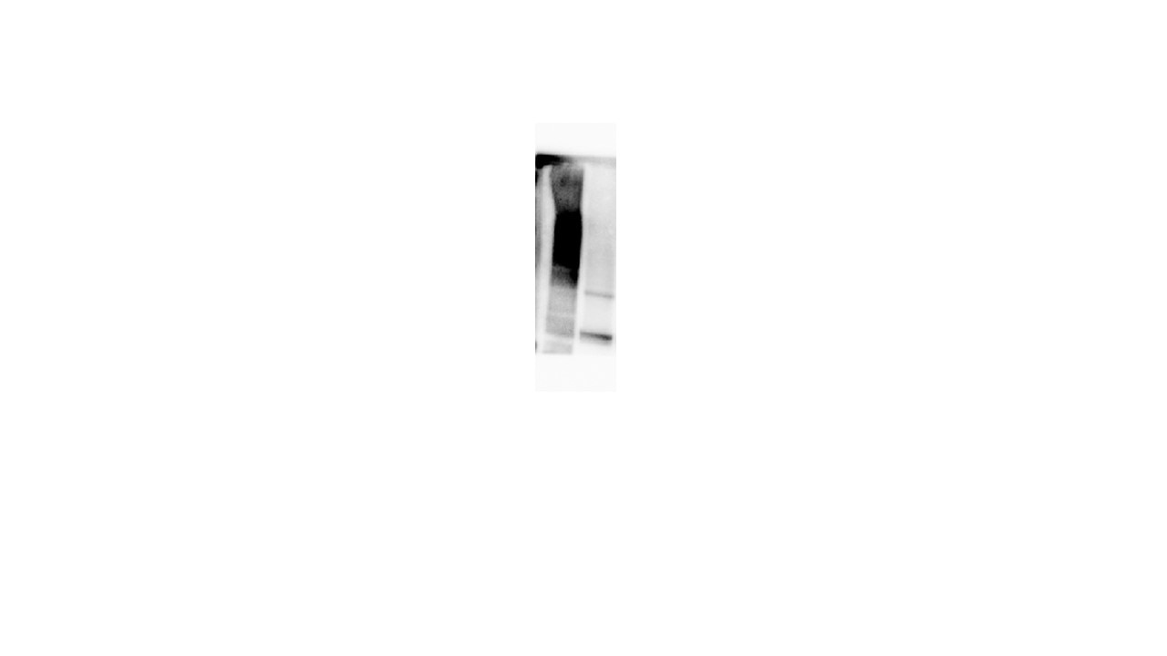
 at dilution of 1:5000 incubated at room temperature for 1.5 hours.")
 with mouse kidney tissue lysate 2160 ug.")
 at dilution of 1:200 (under 40x lens). Heat mediated antigen retrieval with Tris-EDTA buffer (pH 9.0).")
 fixed HEK-293 cells using MFN1 antibody (13798-1-AP) at dilution of 1:400 and CoraLite®488-Conjugated AffiniPure Goat Anti-Rabbit IgG(H+L).")
Applications testées
| Résultats positifs en WB | cellules HSC-T6, cellules T-47D, tissu cérébral de souris, tissu hépatique de souris, tissu rénal de souris |
| Résultats positifs en IP | tissu rénal de souris, |
| Résultats positifs en IHC | tissu rénal humain, il est suggéré de démasquer l'antigène avec un tampon de TE buffer pH 9.0; (*) À défaut, 'le démasquage de l'antigène peut être 'effectué avec un tampon citrate pH 6,0. |
| Résultats positifs en IF/ICC | cellules HEK-293, |
Dilution recommandée
| Application | Dilution |
|---|---|
| Western Blot (WB) | WB : 1:2000-1:10000 |
| Immunoprécipitation (IP) | IP : 0.5-4.0 ug for 1.0-3.0 mg of total protein lysate |
| Immunohistochimie (IHC) | IHC : 1:50-1:500 |
| Immunofluorescence (IF)/ICC | IF/ICC : 1:200-1:800 |
| It is recommended that this reagent should be titrated in each testing system to obtain optimal results. | |
| Sample-dependent, check data in validation data gallery | |
Applications publiées
| KD/KO | See 9 publications below |
| WB | See 321 publications below |
| IHC | See 16 publications below |
| IF | See 20 publications below |
| IP | See 1 publications below |
Informations sur le produit
13798-1-AP cible MFN1 dans les applications de WB, IHC, IF/ICC, IP, ELISA et montre une réactivité avec des échantillons Humain, rat, souris
| Réactivité | Humain, rat, souris |
| Réactivité citée | bovin, canin, Chèvre, Humain, poulet, singe, souris, Hamster, duck |
| Hôte / Isotype | Lapin / IgG |
| Clonalité | Polyclonal |
| Type | Anticorps |
| Immunogène | MFN1 Protéine recombinante Ag4762 |
| Nom complet | mitofusin 1 |
| Masse moléculaire calculée | 741 aa, 84 kDa |
| Poids moléculaire observé | 84 kDa |
| Numéro d’acquisition GenBank | BC040557 |
| Symbole du gène | MFN1 |
| Identification du gène (NCBI) | 55669 |
| Conjugaison | Non conjugué |
| Forme | Liquide |
| Méthode de purification | Purification par affinité contre l'antigène |
| Tampon de stockage | PBS with 0.02% sodium azide and 50% glycerol |
| Conditions de stockage | Stocker à -20°C. Stable pendant un an après l'expédition. L'aliquotage n'est pas nécessaire pour le stockage à -20oC Les 20ul contiennent 0,1% de BSA. |
Informations générales
Mitofusin-1 (MFN1) is a mediator of mitochondrial fusion. This protein and mitofusin 2 are homologs of the Drosophila protein fuzzy onion (Fzo). Mitofusins are large predicted GTPases located in the outer mitochondrial membrane. They are essential for outer membrane fusion by interacting with each other to facilitate mitochondrial targeting. The mitofusins are the first known protein mediator of mitochondrial fusion, and mediate developmentally regulated post-meiotic fusion of mitochondria.Mfn1 is required on adjacent mitochondria to mediate fusion via interactions of a heptad repeat region that mediates oligomerization of the protein(PMID:16892085). Mitofusin 1 and mitofusin 2 are ubiquitinated in a PINK1/parkin-dependent manner upon induction of mitophagy(PMID: 20871098).
Protocole
| Product Specific Protocols | |
|---|---|
| WB protocol for MFN1 antibody 13798-1-AP | Download protocol |
| IHC protocol for MFN1 antibody 13798-1-AP | Download protocol |
| IF protocol for MFN1 antibody 13798-1-AP | Download protocol |
| IP protocol for MFN1 antibody 13798-1-AP | Download protocol |
| Standard Protocols | |
|---|---|
| Click here to view our Standard Protocols |
Publications
| Species | Application | Title |
|---|---|---|
Nature Cellular ATP demand creates metabolically distinct subpopulations of mitochondria
| ||
Cell Res Mitochondria-localized cGAS suppresses ferroptosis to promote cancer progression | ||
Cell Mitocytosis, a migrasome-mediated mitochondrial quality-control process.
| ||
Cell Metab Mitochondrial Dynamics Is Critical for the Full Pluripotency and Embryonic Developmental Potential of Pluripotent Stem Cells. | ||
ACS Cent Sci Macrophage Inactivation by Small Molecule Wedelolactone via Targeting sEH for the Treatment of LPS-Induced Acute Lung Injury |
Avis
The reviews below have been submitted by verified Proteintech customers who received an incentive for providing their feedback.
FH Pierre (Verified Customer) (09-26-2025) | Very good
|
FH Henry (Verified Customer) (09-23-2025) | Good resolution
|
FH S (Verified Customer) (09-23-2024) | Good
 |
FH Ruiting (Verified Customer) (09-18-2020) | Always purchase antibodies from your company, antibody is cheap, and works well. Never has a bad experience in any antibody. Plan to buy two more next week.
|
FH Kishor (Verified Customer) (01-30-2019) | I working good for Western Blotting (1:1000) in human cancer cells but not for rat liver tissue
|

