- Phare
- Validé par KD/KO
Anticorps Polyclonal de lapin anti-MLKL
MLKL Polyclonal Antibody for WB, IHC, IF/ICC, ELISA
Hôte / Isotype
Lapin / IgG
Réactivité testée
Humain et plus (2)
Applications
WB, IHC, IF/ICC, IP, CoIP, ELISA
Conjugaison
Non conjugué
N° de cat : 21066-1-AP
Synonymes
Galerie de données de validation
Applications testées
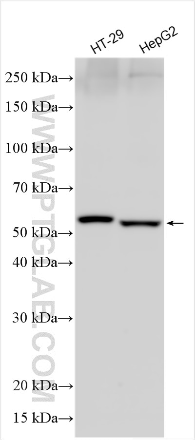
| Résultats positifs en WB | cellules HT-29, cellules HepG2 |
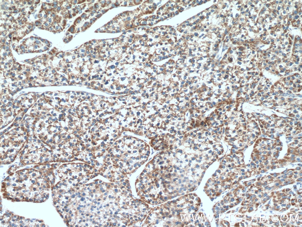
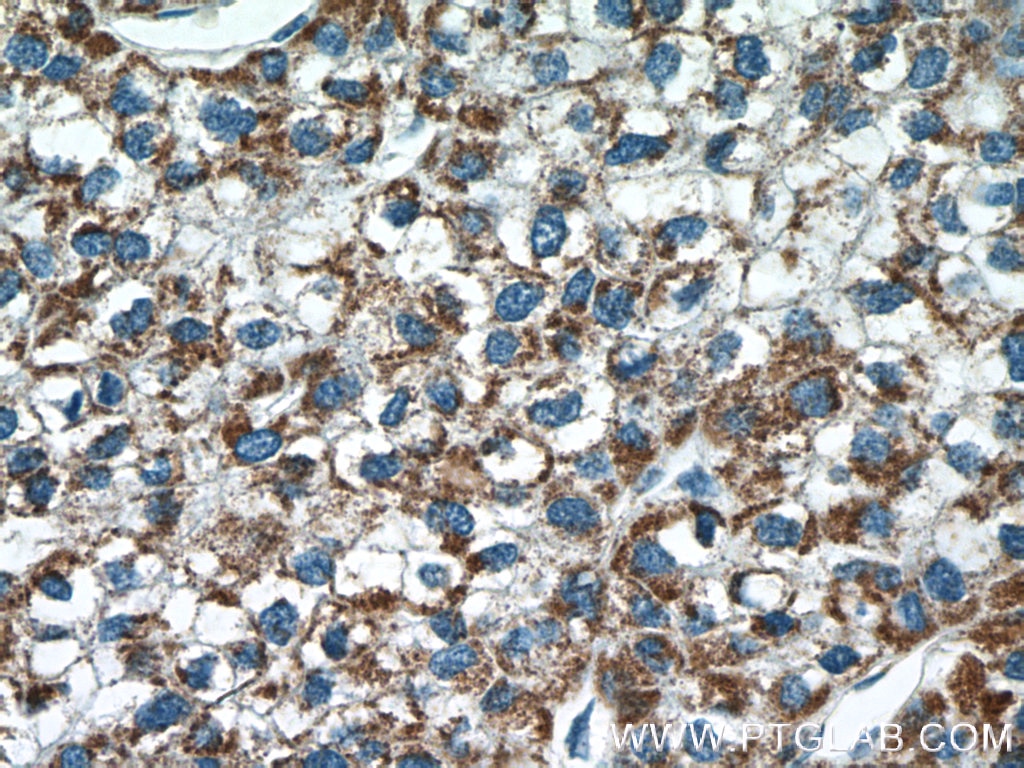
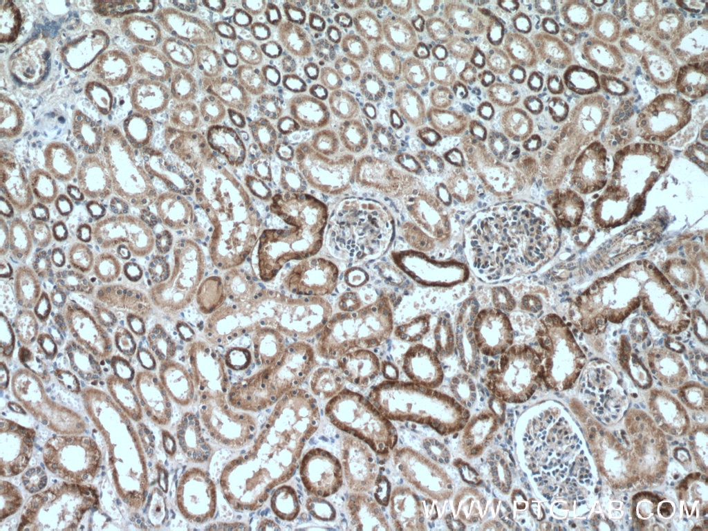
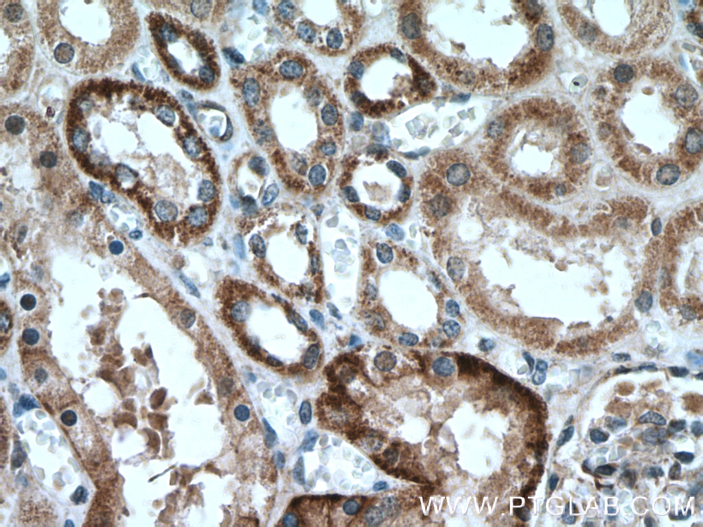
| Résultats positifs en IHC | tissu de cancer du foie humain, tissu rénal humain il est suggéré de démasquer l'antigène avec un tampon de TE buffer pH 9.0; (*) À défaut, 'le démasquage de l'antigène peut être 'effectué avec un tampon citrate pH 6,0. |
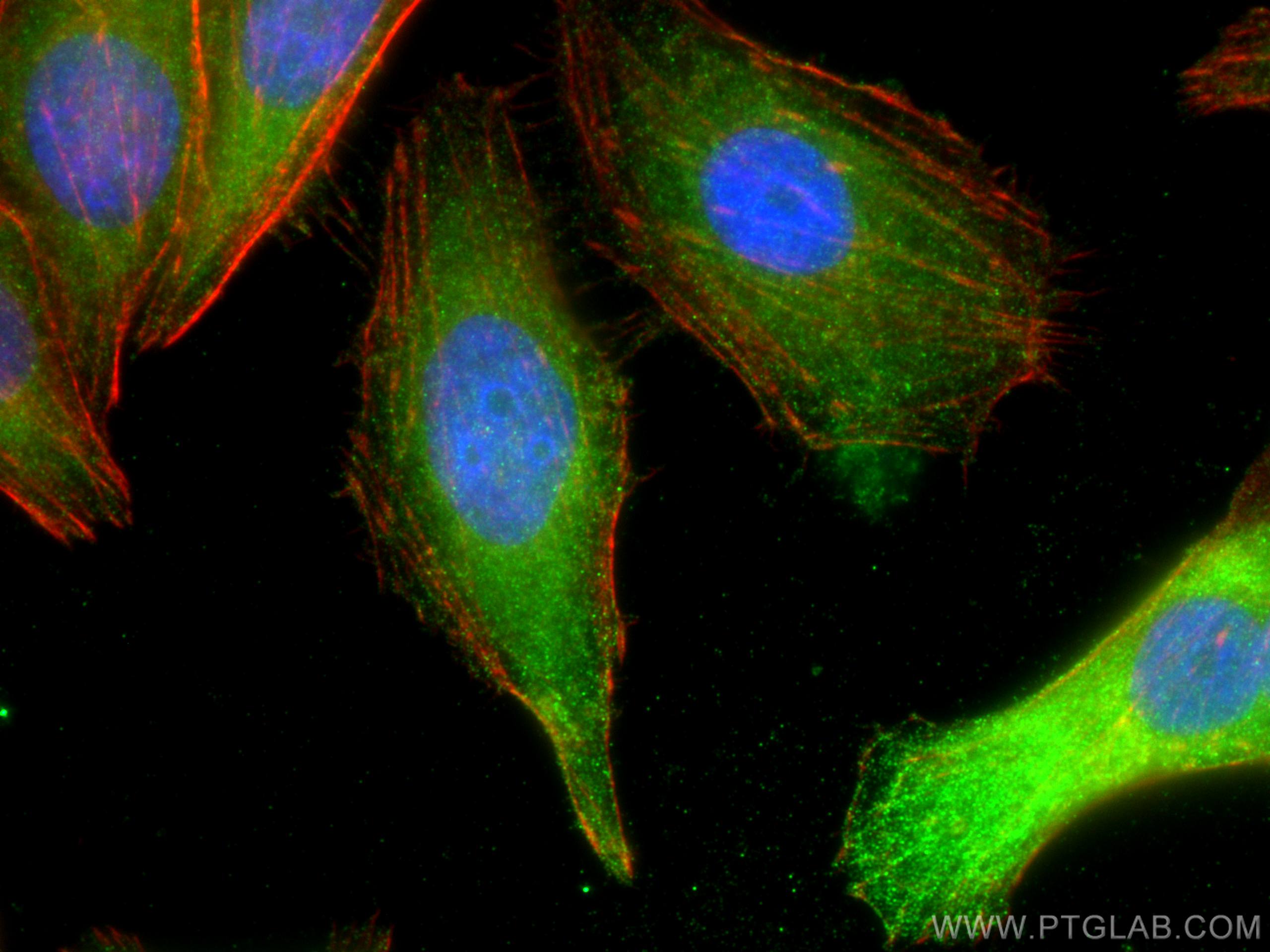
| Résultats positifs en IF/ICC | cellules HepG2, |
Dilution recommandée
| Application | Dilution |
|---|---|
| Western Blot (WB) | WB : 1:500-1:3000 |
| Immunohistochimie (IHC) | IHC : 1:50-1:500 |
| Immunofluorescence (IF)/ICC | IF/ICC : 1:50-1:500 |
| It is recommended that this reagent should be titrated in each testing system to obtain optimal results. | |
| Sample-dependent, check data in validation data gallery | |
Applications publiées
| KD/KO | See 2 publications below |
| WB | See 60 publications below |
| IHC | See 6 publications below |
| IF | See 9 publications below |
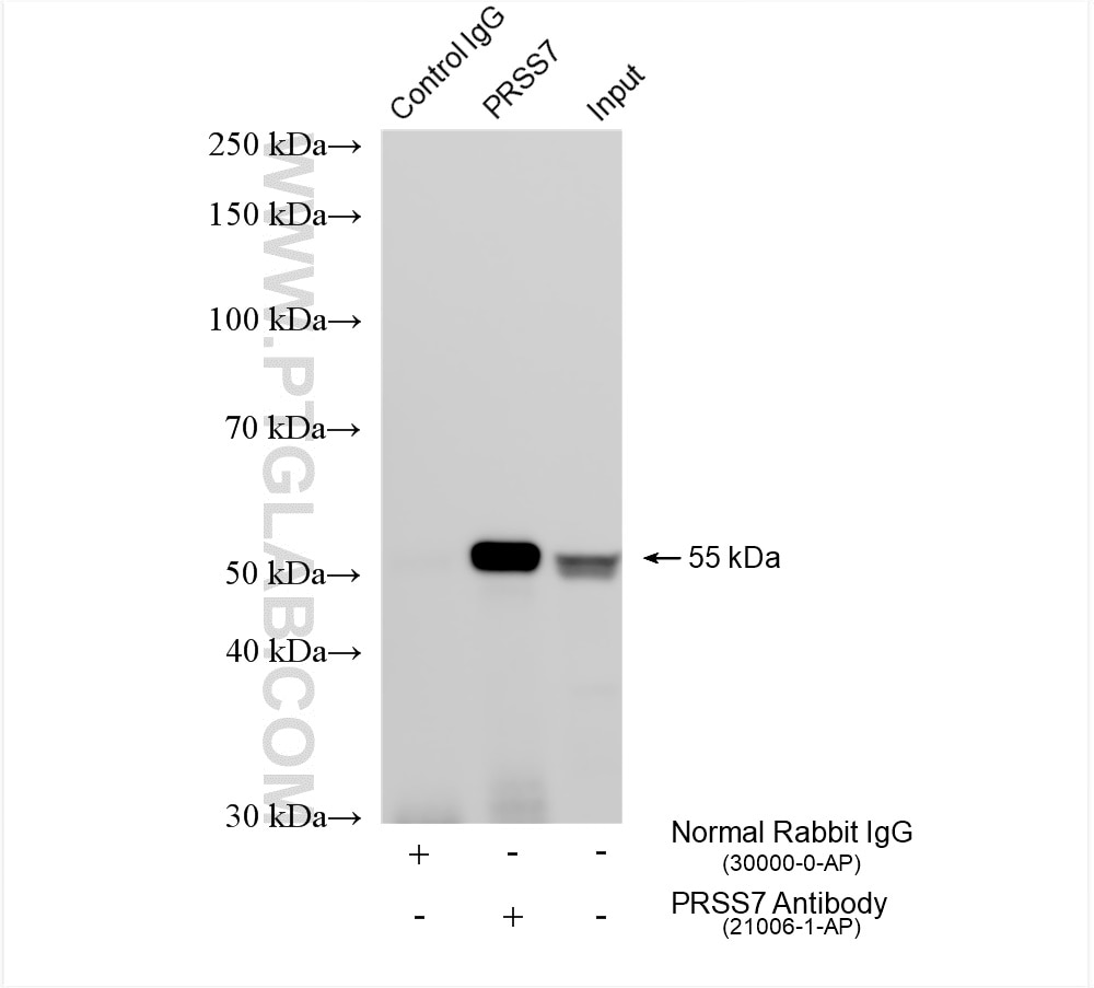
| IP | See 1 publications below |
| CoIP | See 1 publications below |
Informations sur le produit
21066-1-AP cible MLKL dans les applications de WB, IHC, IF/ICC, IP, CoIP, ELISA et montre une réactivité avec des échantillons Humain
| Réactivité | Humain |
| Réactivité citée | rat, Humain, porc |
| Hôte / Isotype | Lapin / IgG |
| Clonalité | Polyclonal |
| Type | Anticorps |
| Immunogène | MLKL Protéine recombinante Ag15188 |
| Nom complet | mixed lineage kinase domain-like |
| Masse moléculaire calculée | 471 aa, 54 kDa |
| Poids moléculaire observé | 54 kDa |
| Numéro d’acquisition GenBank | BC028141 |
| Symbole du gène | MLKL |
| Identification du gène (NCBI) | 197259 |
| Conjugaison | Non conjugué |
| Forme | Liquide |
| Méthode de purification | Purification par affinité contre l'antigène |
| Tampon de stockage | PBS with 0.02% sodium azide and 50% glycerol |
| Conditions de stockage | Stocker à -20°C. Stable pendant un an après l'expédition. L'aliquotage n'est pas nécessaire pour le stockage à -20oC Les 20ul contiennent 0,1% de BSA. |
Informations générales
Mixed lineage kinase domain like pseudokinase (MLKL), belongs to the protein kinase superfamily, has two MW of 54 and 30 kDa. MLKL plays a critical role in tumor necrosis factor (TNF)-induced necroptosis, a programmed cell death process, via interaction with receptor-interacting protein 3 (RIP3), which is a key signaling molecule in necroptosis pathway. High levels of this protein and RIP3 are associated with inflammatory bowel disease in children.
Protocole
| Product Specific Protocols | |
|---|---|
| WB protocol for MLKL antibody 21066-1-AP | Download protocol |
| IHC protocol for MLKL antibody 21066-1-AP | Download protocol |
| IF protocol for MLKL antibody 21066-1-AP | Download protocol |
| IP protocol for MLKL antibody 21066-1-AP | Download protocol |
| Standard Protocols | |
|---|---|
| Click here to view our Standard Protocols |
Publications
| Species | Application | Title |
|---|---|---|
Cell Host Microbe Toxoplasma gondii secreted effectors co-opt host repressor complexes to inhibit necroptosis.
| ||
Cancer Lett Modulation of YBX1-mediated PANoptosis inhibition by PPM1B and USP10 confers chemoresistance to oxaliplatin in gastric cancer | ||
Front Immunol N-acetyl-L-cysteine alleviated the oxidative stress-induced inflammation and necroptosis caused by excessive NiCl2 in primary spleen lymphocytes | ||
Cell Death Discov MLKL deficiency attenuated hepatocyte oxidative DNA damage by activating mitophagy to suppress macrophage cGAS-STING signaling during liver ischemia and reperfusion injury | ||
Cancers (Basel) STAT3β Enhances Sensitivity to Concurrent Chemoradiotherapy by Inducing Cellular Necroptosis in Esophageal Squamous Cell Carcinoma. | ||
Am J Chin Med Neferine, a Bisbenzylisoquinoline Alkaloid, Ameliorates Dextran Sulfate Sodium-Induced Ulcerative Colitis. |
