- Phare
- Validé par KD/KO
Anticorps Polyclonal de lapin anti-MTHFD1L
MTHFD1L Polyclonal Antibody for WB, IHC, IP, ELISA
Hôte / Isotype
Lapin / IgG
Réactivité testée
Humain, rat, souris
Applications
WB, IHC, IP, ELISA
Conjugaison
Non conjugué
N° de cat : 16113-1-AP
Synonymes
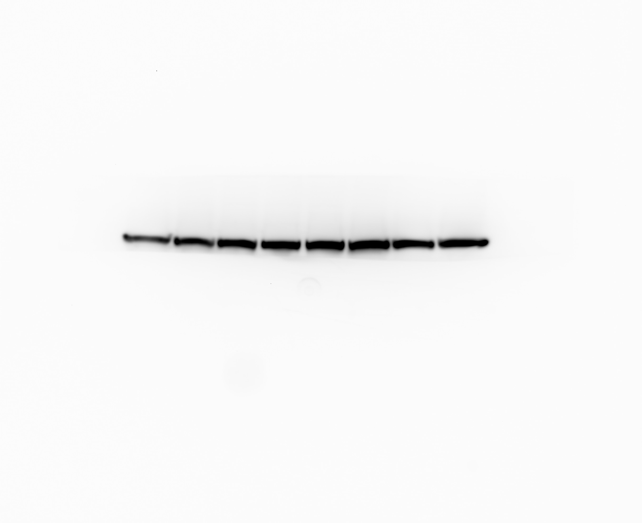
Galerie de données de validation
 at dilution of 1:5000 incubated at room temperature for 1.5 hours.")
 at dilution of 1:500 incubated at room temperature for 1.5 hours.")
 at dilution of 1:1000 incubated at room temperature for 1.5 hours.")
 with HeLa cells lysate 2000ug.")
 at dilution of 1:200 (under 10x lens). Heat mediated antigen retrieval with Tris-EDTA buffer (pH 9.0).")
 at dilution of 1:200 (under 40x lens). Heat mediated antigen retrieval with Tris-EDTA buffer (pH 9.0).")
 at dilution of 1:200 (under 10x lens). Heat mediated antigen retrieval with Tris-EDTA buffer (pH 9.0).")
 at dilution of 1:200 (under 40x lens). Heat mediated antigen retrieval with Tris-EDTA buffer (pH 9.0).")
 fixed HepG2 cells using 16113-1-AP (MTHFD1L antibody) at dilution of 1:100 and CoraLite488-Conjugated AffiniPure Goat Anti-Rabbit IgG(H+L).")
 fixed HepG2 cells using MTHFD1L antibody (16113-1-AP) at dilution of 1:400 and CoraLite®488-Conjugated Goat Anti-Rabbit IgG(H+L) (SA00013-2), CL594-phalloidin (red).")
Applications testées
| Résultats positifs en WB | cellules HEK-293, cellules COLO 320, cellules HeLa, cellules HepG2, tissu ovarien de souris |
| Résultats positifs en IP | cellules HeLa |
| Résultats positifs en IHC | tissu de cancer du foie humain, il est suggéré de démasquer l'antigène avec un tampon de TE buffer pH 9.0; (*) À défaut, 'le démasquage de l'antigène peut être 'effectué avec un tampon citrate pH 6,0. |
Dilution recommandée
| Application | Dilution |
|---|---|
| Western Blot (WB) | WB : 1:2000-1:10000 |
| Immunoprécipitation (IP) | IP : 0.5-4.0 ug for 1.0-3.0 mg of total protein lysate |
| Immunohistochimie (IHC) | IHC : 1:50-1:500 |
| It is recommended that this reagent should be titrated in each testing system to obtain optimal results. | |
| Sample-dependent, check data in validation data gallery | |
Applications publiées
| KD/KO | See 8 publications below |
| WB | See 18 publications below |
| IHC | See 2 publications below |
Informations sur le produit
16113-1-AP cible MTHFD1L dans les applications de WB, IHC, IP, ELISA et montre une réactivité avec des échantillons Humain, rat, souris
| Réactivité | Humain, rat, souris |
| Réactivité citée | Humain, souris |
| Hôte / Isotype | Lapin / IgG |
| Clonalité | Polyclonal |
| Type | Anticorps |
| Immunogène | MTHFD1L Protéine recombinante Ag9036 |
| Nom complet | methylenetetrahydrofolate dehydrogenase (NADP+ dependent) 1-like |
| Masse moléculaire calculée | 978 aa, 106 kDa |
| Poids moléculaire observé | 106 kDa |
| Numéro d’acquisition GenBank | BC017477 |
| Symbole du gène | MTHFD1L |
| Identification du gène (NCBI) | 25902 |
| Conjugaison | Non conjugué |
| Forme | Liquide |
| Méthode de purification | Purification par affinité contre l'antigène |
| Tampon de stockage | PBS with 0.02% sodium azide and 50% glycerol |
| Conditions de stockage | Stocker à -20°C. Stable pendant un an après l'expédition. L'aliquotage n'est pas nécessaire pour le stockage à -20oC Les 20ul contiennent 0,1% de BSA. |
Informations générales
MTHFD1L(Monofunctional C1-tetrahydrofolate synthase, mitochondrial) is also named as FTHFSDC1(Formyltetrahydrofolate synthetase). MTHFD1L enzyme is present in mitochondria from normal embryonic tissues and embryonic fibroblast cell lines, and embryonic mitochondria possess the ability to synthesize formate from glycine. It catalyzes the final step in the mitochondrial conversion of 1-C units to formate in embryos. Moreover, MTHFD1L levels were substantially higher in embryonic mitochondria than in adult liver mitochondria and embryonic mitochondria exhibited greater formate production(PMID:19948730). It has 2 isoforms produced by alternative splicing.
Protocole
| Product Specific Protocols | |
|---|---|
| WB protocol for MTHFD1L antibody 16113-1-AP | Download protocol |
| IHC protocol for MTHFD1L antibody 16113-1-AP | Download protocol |
| IF protocol for MTHFD1L antibody 16113-1-AP | Download protocol |
| IP protocol for MTHFD1L antibody 16113-1-AP | Download protocol |
| Standard Protocols | |
|---|---|
| Click here to view our Standard Protocols |
Publications
| Species | Application | Title |
|---|---|---|
Cell Metab Fibroblast Growth Factor 21 Drives Dynamics of Local and Systemic Stress Responses in Mitochondrial Myopathy with mtDNA Deletions. | ||
Cell Metab Tumor Reliance on Cytosolic versus Mitochondrial One-Carbon Flux Depends on Folate Availability. | ||
Cell Metab Mitochondrial DNA Replication Defects Disturb Cellular dNTP Pools and Remodel One-Carbon Metabolism. | ||
J Pineal Res Melatonin modulates metabolic remodeling in HNSCC by suppressing MTHFD1L-formate axis.
| ||
Avis
The reviews below have been submitted by verified Proteintech customers who received an incentive for providing their feedback.
FH Furkan Emre (Verified Customer) (02-21-2022) | I am satisfied with the antibody. It worked as I was expecting from it. I would buy it again. I also add a figure showing chemiluminescence detection of MTHFD1L using your antibody.
 |

