- Phare
- Validé par KD/KO
Anticorps Polyclonal de lapin anti-MYL9
MYL9 Polyclonal Antibody for WB, IHC, IF/ICC, IF-P, ELISA
Hôte / Isotype
Lapin / IgG
Réactivité testée
Humain, rat, souris
Applications
WB, IHC, IF/ICC, IF-P, IP, ELISA
Conjugaison
Non conjugué
N° de cat : 15354-1-AP
Synonymes
Galerie de données de validation
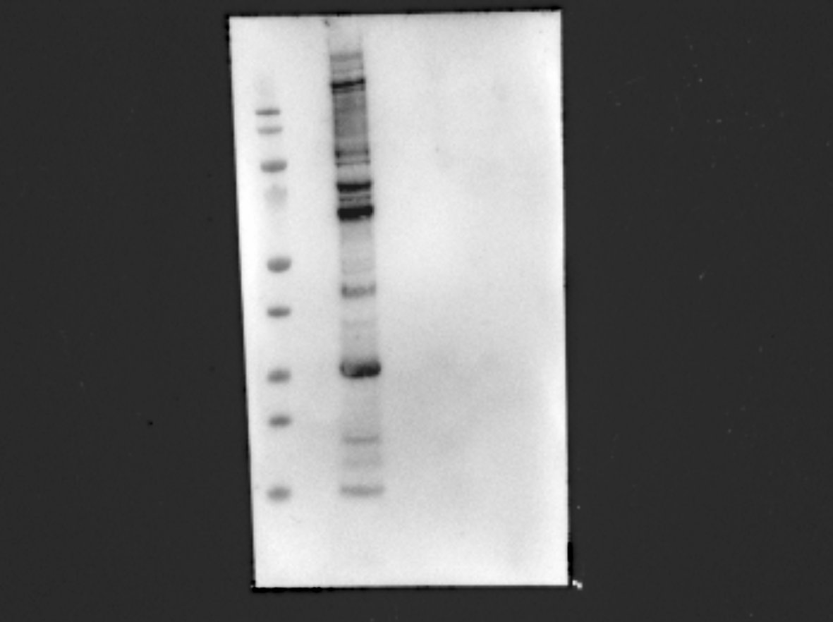
 at dilution of 1:3000 incubated at room temperature for 1.5 hours.")
 at dilution of 1:600 incubated at room temperature for 1.5 hours.")
 at dilution of 1:500 (under 40x lens. Heat mediated antigen retrieval with Tris-EDTA buffer (pH 9.0).")
 at dilution of 1:500 (under 10x lens. Heat mediated antigen retrieval with Tris-EDTA buffer (pH 9.0).")
 at dilution of 1:500 (under 40x lens. Heat mediated antigen retrieval with Tris-EDTA buffer (pH 9.0).")
 at dilution of 1:500 (under 10x lens. Heat mediated antigen retrieval with Tris-EDTA buffer (pH 9.0).")
 at dilution of 1:500 (under 10x lens. Heat mediated antigen retrieval with Tris-EDTA buffer (pH 9.0).")
 fixed paraffin-embedded mouse heart tissue using MYL9 antibody (15354-1-AP) at dilution of 1:200 and CoraLite®488-Conjugated Goat Anti-Rabbit IgG(H+L) (SA00013-2). Heat mediated antigen retrieval with Tris-EDTA buffer (pH 9.0).")
 fixed paraffin-embedded mouse heart tissue using MYL9 antibody (15354-1-AP) at dilution of 1:200 and CoraLite®488-Conjugated Goat Anti-Rabbit IgG(H+L) (SA00013-2). Heat mediated antigen retrieval with Tris-EDTA buffer (pH 9.0).")
 fixed MCF-7 cells using MYL9 antibody (15354-1-AP) at dilution of 1:400 and CoraLite®488-Conjugated AffiniPure Goat Anti-Rabbit IgG(H+L).")
 fixed C2C12 cells using MYL9 antibody (15354-1-AP) at dilution of 1:400 and CoraLite®488-Conjugated AffiniPure Goat Anti-Rabbit IgG(H+L).")
.")
Applications testées
| Résultats positifs en WB | tissu de côlon de souris, cellules Caco-2, tissu de côlon de rat |
| Résultats positifs en IHC | tissu de cancer du foie humain, tissu de cancer du côlon humain il est suggéré de démasquer l'antigène avec un tampon de TE buffer pH 9.0; (*) À défaut, 'le démasquage de l'antigène peut être 'effectué avec un tampon citrate pH 6,0. |
| Résultats positifs en IF-P | tissu cardiaque de souris, |
| Résultats positifs en IF/ICC | cellules MCF-7, cellules C2C12 |
Dilution recommandée
| Application | Dilution |
|---|---|
| Western Blot (WB) | WB : 1:500-1:5000 |
| Immunohistochimie (IHC) | IHC : 1:250-1:1000 |
| Immunofluorescence (IF)-P | IF-P : 1:50-1:500 |
| Immunofluorescence (IF)/ICC | IF/ICC : 1:200-1:800 |
| It is recommended that this reagent should be titrated in each testing system to obtain optimal results. | |
| Sample-dependent, check data in validation data gallery | |
Applications publiées
| KD/KO | See 1 publications below |
| WB | See 9 publications below |
| IHC | See 2 publications below |
| IF | See 3 publications below |
| IP | See 1 publications below |
Informations sur le produit
15354-1-AP cible MYL9 dans les applications de WB, IHC, IF/ICC, IF-P, IP, ELISA et montre une réactivité avec des échantillons Humain, rat, souris
| Réactivité | Humain, rat, souris |
| Réactivité citée | rat, Humain, souris |
| Hôte / Isotype | Lapin / IgG |
| Clonalité | Polyclonal |
| Type | Anticorps |
| Immunogène | MYL9 Protéine recombinante Ag7600 |
| Nom complet | myosin, light chain 9, regulatory |
| Masse moléculaire calculée | 20 kDa |
| Poids moléculaire observé | 20 kDa |
| Numéro d’acquisition GenBank | BC002648 |
| Symbole du gène | MYL9 |
| Identification du gène (NCBI) | 10398 |
| Conjugaison | Non conjugué |
| Forme | Liquide |
| Méthode de purification | Purification par affinité contre l'antigène |
| Tampon de stockage | PBS with 0.02% sodium azide and 50% glycerol |
| Conditions de stockage | Stocker à -20°C. Stable pendant un an après l'expédition. L'aliquotage n'est pas nécessaire pour le stockage à -20oC Les 20ul contiennent 0,1% de BSA. |
Informations générales
Myosin regulatory light polypeptide 9 (MYL9), also known as MLC2, belongs to the myosin regulatory subunits. It plays an important role in regulation of both smooth muscle and nonmuscle cell contractile activity via its phosphorylation at Thr19 and Ser20. Implicated in cytokinesis, receptor capping, and cell locomotion (PMID:11942626, PMID:2526655). Some studies have demonstrated that MYL9 may play important roles in various human cancers. The expression and phosphorylation of MYL9 (Thr19/Ser20) may be increased in human breast (PMID: 22144583) and liver cancers (PMID: 18648664), while decreased in human colon (PMID: 22752057) and bladder cancers (PMID: 21139803). MYL9 was the only gene differentially expressed in the aged versus young injured arteries in the rat smooth muscle cell layers (PMID:22003410).
Protocole
| Product Specific Protocols | |
|---|---|
| WB protocol for MYL9 antibody 15354-1-AP | Download protocol |
| IHC protocol for MYL9 antibody 15354-1-AP | Download protocol |
| IF protocol for MYL9 antibody 15354-1-AP | Download protocol |
| Standard Protocols | |
|---|---|
| Click here to view our Standard Protocols |
Publications
| Species | Application | Title |
|---|---|---|
J Biol Chem Brain-Derived Neurotrophic Factor regulates LYN kinase mediated Myosin Light Chain Kinase activation to modulate non-muscle myosin II activity in hippocampal neurons. | ||
Sci Rep Proteomic profiling of concurrently isolated primary microvascular endothelial cells, pericytes, and vascular smooth muscle cells from adult mouse heart. | ||
Front Med (Lausanne) Depiction of Aging-Based Molecular Phenotypes With Diverse Clinical Prognosis and Immunological Features in Gastric Cancer.
| ||
Oncol Rep Fenretinide inhibits the proliferation and migration of human liver cancer HepG2 cells by downregulating the activation of myosin light chain kinase through the p38‑MAPK signaling pathway. | ||
JCI Insight Transient expansion and myofibroblast conversion of adipogenic lineage precursors mediate bone marrow repair after radiation. | ||
Front Cell Dev Biol Paracrine HB-EGF signaling reduce enhanced contractile and energetic state of activated decidual fibroblasts by rebalancing SRF-MRTF-TCF transcriptional axis |
Avis
The reviews below have been submitted by verified Proteintech customers who received an incentive for providing their feedback.
FH Irem (Verified Customer) (05-18-2022) | 20 kDa band can be observed but there are other unspesific bands. Marker: Pageruler protein prestained marker
 |

