- Phare
- Validé par KD/KO
Anticorps Polyclonal de lapin anti-MYO6
MYO6 Polyclonal Antibody for WB, IHC, IF/ICC, IF-P, IP, ELISA
Hôte / Isotype
Lapin / IgG
Réactivité testée
Humain, souris
Applications
WB, IHC, IF/ICC, IF-P, IP, ELISA
Conjugaison
Non conjugué
N° de cat : 26778-1-AP
Synonymes
Galerie de données de validation
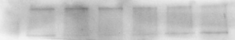
 at dilution of 1:500 incubated at room temperature for 1.5 hours.")
 at dilution of 1:15000 incubated at room temperature for 1.5 hours.")
 with PC-3 cells lysate 2000 ug.")
 at dilution of 1:200 (under 40x lens). Heat mediated antigen retrieval with Tris-EDTA buffer (pH 9.0).")
 at dilution of 1:200 (under 40x lens).")
 fixed mouse small intestine tissue using 26778-1-AP (MYO6 antibody) at dilution of 1:50 and Alexa Fluor 488-Conjugated AffiniPure Goat Anti-Rabbit IgG(H+L).")
 fixed MCF-7 cells using MYO6 antibody (26778-1-AP) at dilution of 1:400 and Multi-rAb CoraLite ® Plus 488-Goat Anti-Rabbit Recombinant Secondary Antibody (H+L) (RGAR002).")
Applications testées
| Résultats positifs en WB | cellules HEK-293, cellules DU 145, cellules LNCaP, cellules MCF-7, tissu d'intestin grêle de souris |
| Résultats positifs en IP | cellules PC-3, |
| Résultats positifs en IHC | tissu de cancer de la prostate humain, tissu d'intestin grêle humain il est suggéré de démasquer l'antigène avec un tampon de TE buffer pH 9.0; (*) À défaut, 'le démasquage de l'antigène peut être 'effectué avec un tampon citrate pH 6,0. |
| Résultats positifs en IF-P | tissu d'intestin grêle de souris, |
| Résultats positifs en IF/ICC | cellules MCF-7, |
Dilution recommandée
| Application | Dilution |
|---|---|
| Western Blot (WB) | WB : 1:5000-1:50000 |
| Immunoprécipitation (IP) | IP : 0.5-4.0 ug for 1.0-3.0 mg of total protein lysate |
| Immunohistochimie (IHC) | IHC : 1:50-1:500 |
| Immunofluorescence (IF)-P | IF-P : 1:50-1:500 |
| Immunofluorescence (IF)/ICC | IF/ICC : 1:200-1:800 |
| It is recommended that this reagent should be titrated in each testing system to obtain optimal results. | |
| Sample-dependent, check data in validation data gallery | |
Applications publiées
| KD/KO | See 1 publications below |
| WB | See 4 publications below |
| IHC | See 3 publications below |
| IF | See 3 publications below |
Informations sur le produit
26778-1-AP cible MYO6 dans les applications de WB, IHC, IF/ICC, IF-P, IP, ELISA et montre une réactivité avec des échantillons Humain, souris
| Réactivité | Humain, souris |
| Réactivité citée | Humain, souris |
| Hôte / Isotype | Lapin / IgG |
| Clonalité | Polyclonal |
| Type | Anticorps |
| Immunogène | MYO6 Protéine recombinante Ag24906 |
| Nom complet | myosin VI |
| Masse moléculaire calculée | 1285 aa, 149 kDa |
| Poids moléculaire observé | 145-150 kDa |
| Numéro d’acquisition GenBank | BC146764 |
| Symbole du gène | MYO6 |
| Identification du gène (NCBI) | 4646 |
| Conjugaison | Non conjugué |
| Forme | Liquide |
| Méthode de purification | Purification par affinité contre l'antigène |
| Tampon de stockage | PBS with 0.02% sodium azide and 50% glycerol |
| Conditions de stockage | Stocker à -20°C. Stable pendant un an après l'expédition. L'aliquotage n'est pas nécessaire pour le stockage à -20oC Les 20ul contiennent 0,1% de BSA. |
Informations générales
MYO6, an actin-based motor protein, is the only myosin known to move toward the minus end of actin filaments. MYO6 is highly expressed in the inner and outer hair cells of the ear, retina, and polarized epithelial cells such as kidney proximal tubule cells and intestinal enterocytes. And it participates in a wide range of biological processes within cells, including clathrin-mediated endocytosis, vesicular membrane traffic, polarized secretion, and autophagy (PMID: 23620821; PMID: 28591580). Previous studies showed that MYO6 is upregulated in various types of cancer, and it has been widely reported to contribute to tumor cell migration and metastasis. Some articles indicate that MYO6 is associated with prostate cancer, lung cancer, human colorectal cancer and gastric cancer (PMID: 29022908).
Protocole
| Product Specific Protocols | |
|---|---|
| WB protocol for MYO6 antibody 26778-1-AP | Download protocol |
| IHC protocol for MYO6 antibody 26778-1-AP | Download protocol |
| IF protocol for MYO6 antibody 26778-1-AP | Download protocol |
| IP protocol for MYO6 antibody 26778-1-AP | Download protocol |
| Standard Protocols | |
|---|---|
| Click here to view our Standard Protocols |
Publications
| Species | Application | Title |
|---|---|---|
Nat Commun Structure of Myosin VI/Tom1 complex reveals a cargo recognition mode of Myosin VI for tethering. | ||
Cell Signal Elevated expression of myosin VI contributes to breast cancer progression via MAPK/ERK signaling pathway
| ||
Nat Commun MYL3 protects chondrocytes from senescence by inhibiting clathrin-mediated endocytosis and activating of Notch signaling | ||
Int J Toxicol Copper Chaperone Atox1 Protected the Cochlea From Cisplatin by Regulating the Copper Transport Family and Cell Cycle | ||
Int J Mol Sci The Suppression of the Epithelial to Mesenchymal Transition in Prostate Cancer through the Targeting of MYO6 Using MiR-145-5p | ||
Cancer Manag Res The Actin Motor Protein Myosin 6 Contributes to Cell Migration and Expression of GIPC1 and Septins in Breast Cancer Cells |
Avis
The reviews below have been submitted by verified Proteintech customers who received an incentive for providing their feedback.
FH Wojciech (Verified Customer) (04-20-2023) | We used this antibody to assess protein levels in human T cells. We got a good signal with 1:1000 dilution with observed molecular weight at ~150kDa.
|
FH Sara (Verified Customer) (10-25-2020) | B cells (mouse) in RIPA buffer. 10 µg of protein per lane were loaded on a 10% PAGE-SDS gel. Transfer is not optimised for high MW proteins, so I could get a better blot. Two bands are detected, one of 150 kDa and one > 200 kDa.
 |

