Anticorps Polyclonal de lapin anti-IBA1
IBA1 Polyclonal Antibody for Single Cell (Intra)
Hôte / Isotype
Lapin / IgG
Réactivité testée
Humain
Applications
Single Cell (Intra)
Conjugaison
5CFLX Fluorescent Dye
N° de cat : G10904-1-5C
Synonymes
Galerie de données de validation
Applications testées
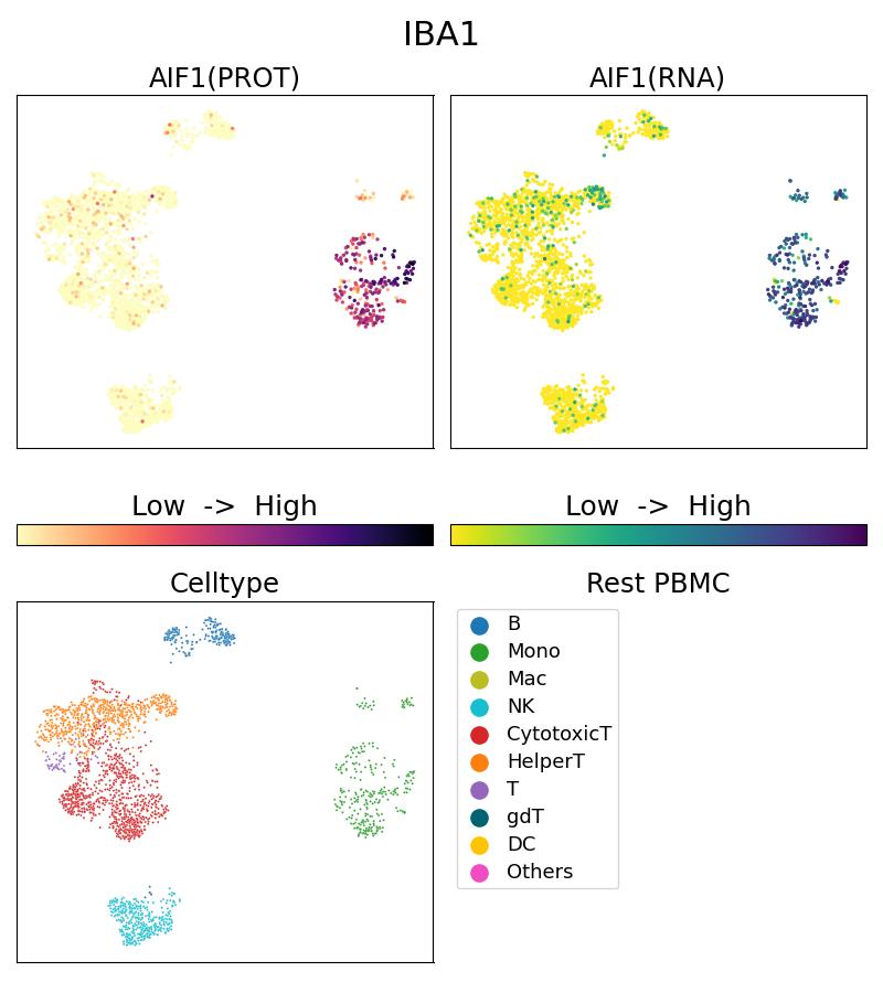
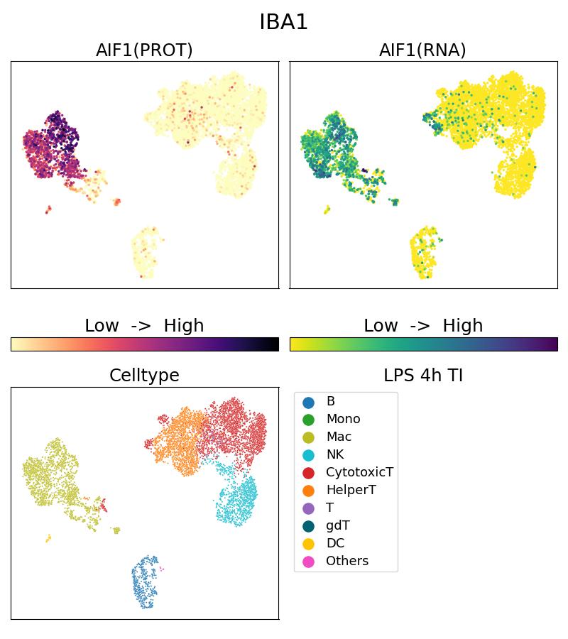
| Résultats positifs en Single Cell (Intra) | 10x Genomics Gene Expression Flex with Feature Barcodes and Multiplexing product. |
Dilution recommandée
| Application | Dilution |
|---|---|
| SINGLE CELL (INTRA) | SINGLE CELL (INTRA) : <0.5ug/test |
| It is recommended that this reagent should be titrated in each testing system to obtain optimal results. | |
| Sample-dependent, check data in validation data gallery | |
Informations sur le produit
G10904-1-5C cible IBA1 dans les applications de Single Cell (Intra) et montre une réactivité avec des échantillons Humain
| Réactivité | Humain |
| Hôte / Isotype | Lapin / IgG |
| Clonalité | Polyclonal |
| Type | Anticorps |
| Immunogène | IBA1 Protéine recombinante Ag1363 |
| Nom complet | allograft inflammatory factor 1 |
| Masse moléculaire calculée | 17 kDa |
| Numéro d’acquisition GenBank | BC009474 |
| Symbole du gène | IBA1 |
| Identification du gène (NCBI) | 199 |
| Conjugaison | 5CFLX Fluorescent Dye |
| Forme | Liquide |
| Méthode de purification | |
| Tampon de stockage | PBS with 1mM EDTA and 0.09% sodium azide |
| Conditions de stockage | 2-8°C Stable for one year after shipment. 20ul contiennent 0,1% de BSA. |
Informations générales
What is the molecular weight of IBA1?
The molecular weight of IBA1 is 16.7 kD.
What is IBA1?
Ionized calcium-binding adaptor molecule 1 (IBA1), also known as Allograft inflammatory factor-1 (AIF-1), is an inflammation-responsive scaffold protein expressed and secreted from macrophages. Microglia response factor (MRF-1) and daintain are also similar, and likely identical, proteins (PMIDs: 29749461, 9630473, 23792284).
What is the function of IBA1?
IBA1 is necessary for macrophage survival, and it is also a key molecule in proinflammatory activity (PMID: 29749461).
Where is IBA1 localized?
IBA1 is a cytoplasmic protein, often expressed in immunocytes, macrophages, and microglia. It can be used as a marker for normal (not 'dark') microglia in brain tissue, as IBA1 is expressed by all microglial cell subpopulations (PMIDs: 29749461, 11943136, 26847266, 9630473).
Is IBA1 upregulated in active immunophages?
Yes; its expression is associated with inflammatory activity (PMID: 29749461).
How do IBA1-positive microglia differ from microglia that express less or no IBA1?
'Dark' microglia is a recently described phenotype associated with Alzheimer's disease pathology and chronic stress; these dark microglia express decreased IBA1 (often punctiform), and show distinct ultrastructural differences from 'normal' microglia as well as condensed and electron-dense cytoplasm and nucleoplasm. Normal microglia generally display strong, diffuse expression of IBA1. IBA1-positive microglial processes in normal conditions also have a strong tendency to exclusively contact synaptic elements, such as axon terminals, dendritic spines, and synaptic clefts (PMIDs: 29992181, 26847266).
How is IBA1 related to Alzheimer's disease and pain?
Exacerbated immunoactivity of IBA1, particularly in proximity to amyloid plaques, is prominently featured in AD pathology. IBA1, along with other microglial markers, are also associated with pain, and robust microglial reactions often follow spinal cord injury (PMIDs: 29992181, 23792284).


