- Phare
- Validé par KD/KO
Anticorps Polyclonal de lapin anti-NLRP3
NLRP3 Polyclonal Antibody for WB, IHC, IF/ICC, FC (Intra), IP, ELISA
Hôte / Isotype
Lapin / IgG
Réactivité testée
Humain et plus (3)
Applications
WB, IHC, IF/ICC, FC (Intra), IP, CoIP, ELISA, Cell treatment
Conjugaison
Non conjugué
N° de cat : 19771-1-AP
Synonymes
Galerie de données de validation
Applications testées
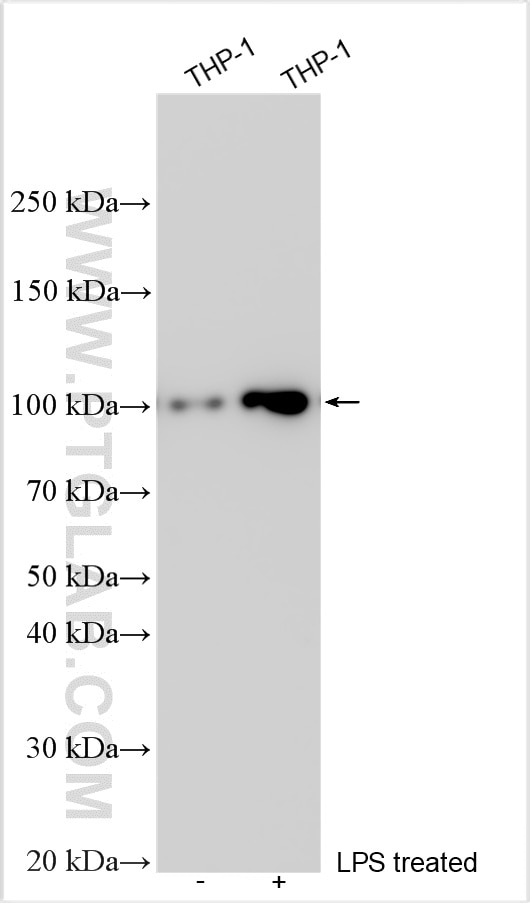
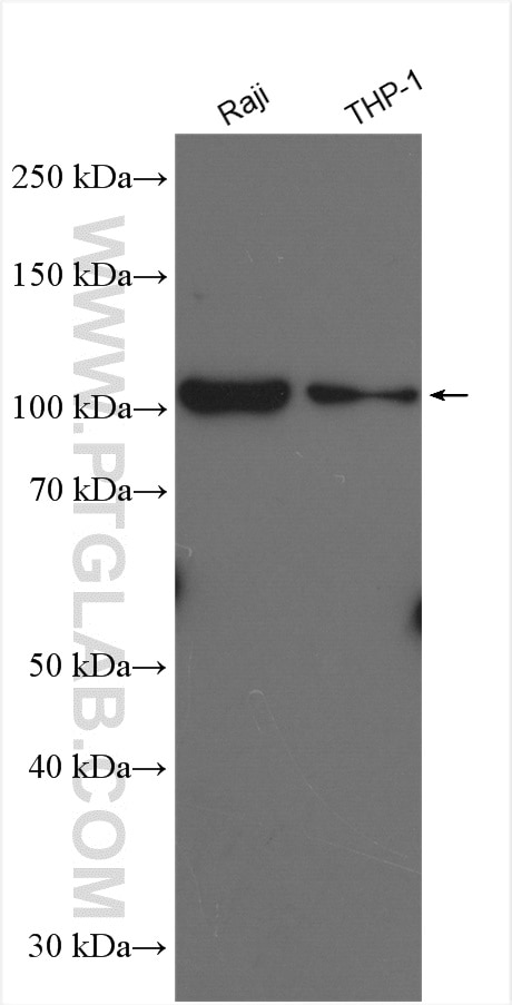
| Résultats positifs en WB | cellules THP-1, cellules Raji, cellules THP-1 traitées au LPS |
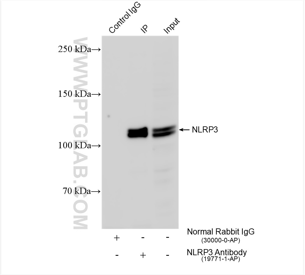
| Résultats positifs en IP | cellules THP-1, |
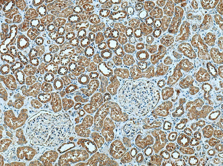
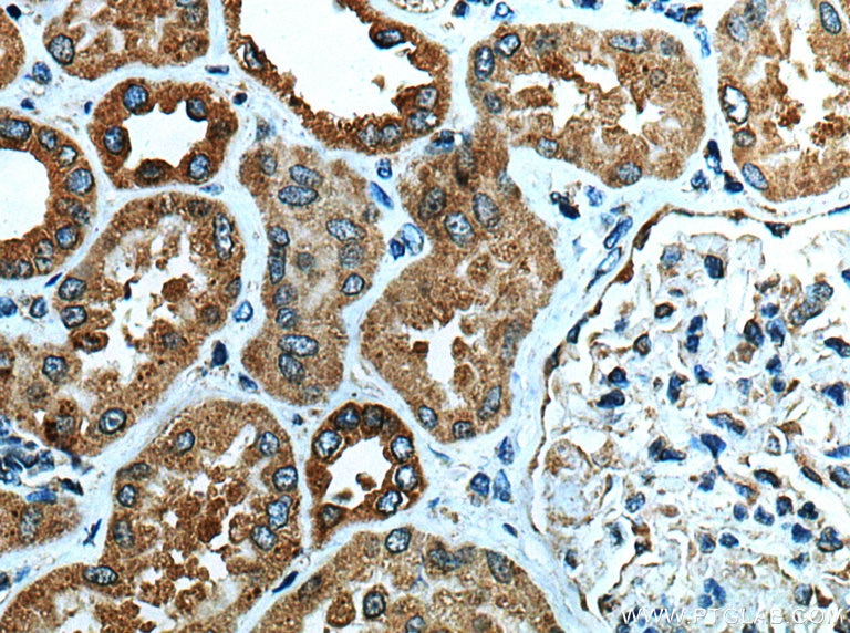
| Résultats positifs en IHC | tissu rénal humain, il est suggéré de démasquer l'antigène avec un tampon de TE buffer pH 9.0; (*) À défaut, 'le démasquage de l'antigène peut être 'effectué avec un tampon citrate pH 6,0. |
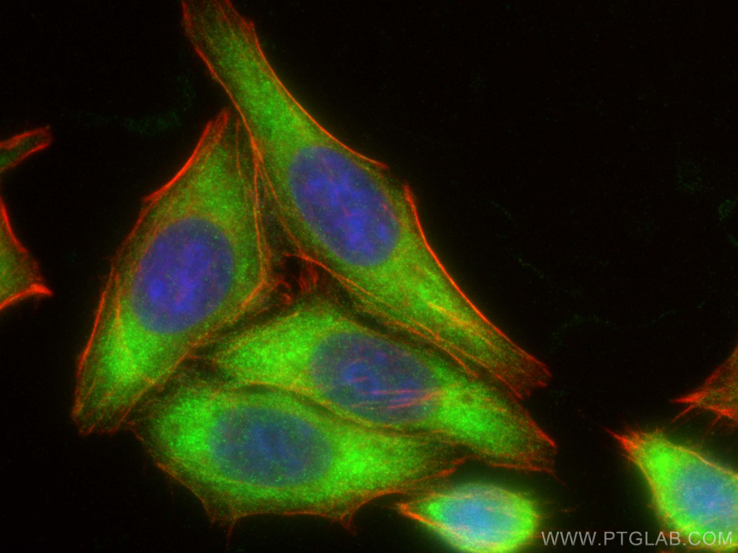
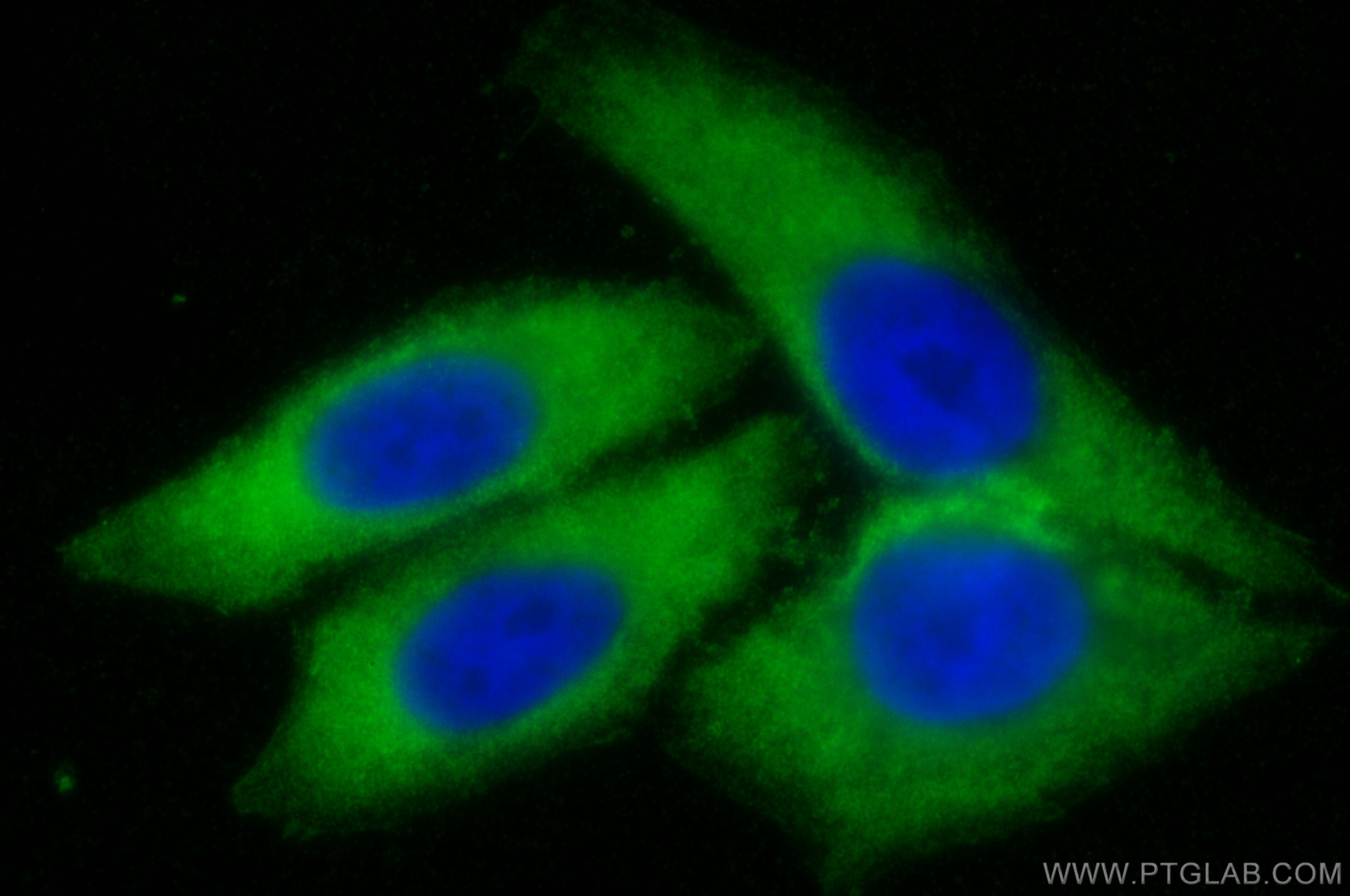
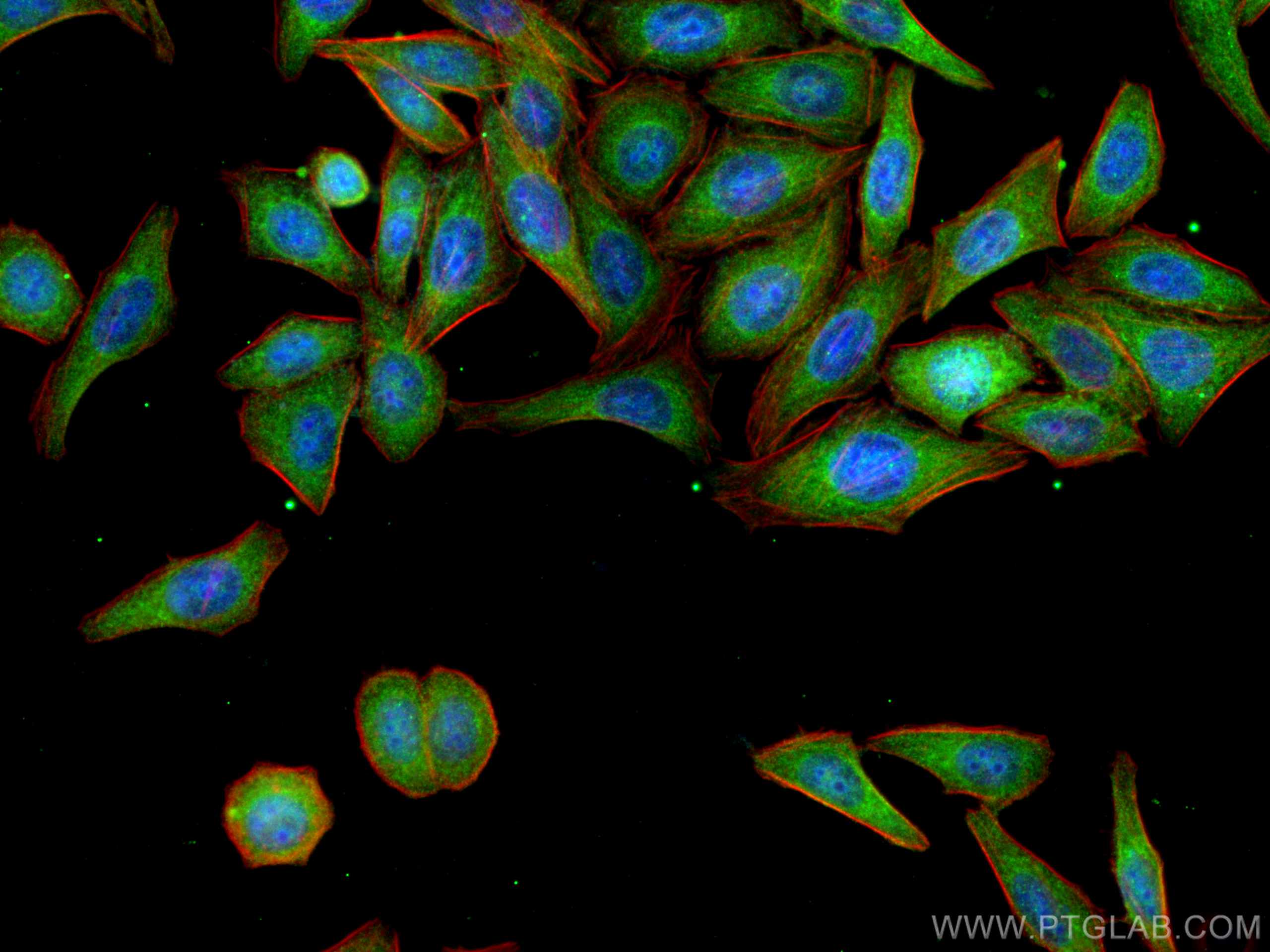
| Résultats positifs en IF/ICC | cellules HepG2, |
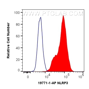
| Résultats positifs en FC (Intra) | cellules THP-1 |
Dilution recommandée
| Application | Dilution |
|---|---|
| Western Blot (WB) | WB : 1:500-1:2000 |
| Immunoprécipitation (IP) | IP : 0.5-4.0 ug for 1.0-3.0 mg of total protein lysate |
| Immunohistochimie (IHC) | IHC : 1:50-1:500 |
| Immunofluorescence (IF)/ICC | IF/ICC : 1:50-1:500 |
| Flow Cytometry (FC) (INTRA) | FC (INTRA) : 0.40 ug per 10^6 cells in a 100 µl suspension |
| It is recommended that this reagent should be titrated in each testing system to obtain optimal results. | |
| Sample-dependent, check data in validation data gallery | |
Applications publiées
| KD/KO | See 19 publications below |
| WB | See 507 publications below |
| IHC | See 120 publications below |
| IF | See 146 publications below |
| IP | See 9 publications below |
| ELISA | See 1 publications below |
| CoIP | See 7 publications below |
Informations sur le produit
19771-1-AP cible NLRP3 dans les applications de WB, IHC, IF/ICC, FC (Intra), IP, CoIP, ELISA, Cell treatment et montre une réactivité avec des échantillons Humain
| Réactivité | Humain |
| Réactivité citée | bovin, Chèvre, Humain, porc |
| Hôte / Isotype | Lapin / IgG |
| Clonalité | Polyclonal |
| Type | Anticorps |
| Immunogène | Peptide |
| Nom complet | NLR family, pyrin domain containing 3 |
| Masse moléculaire calculée | 118 kDa |
| Poids moléculaire observé | 110 kDa |
| Numéro d’acquisition GenBank | NM_001127461 |
| Symbole du gène | NLRP3 |
| Identification du gène (NCBI) | 114548 |
| Conjugaison | Non conjugué |
| Forme | Liquide |
| Méthode de purification | Purification par affinité contre l'antigène |
| Tampon de stockage | PBS with 0.02% sodium azide and 50% glycerol |
| Conditions de stockage | Stocker à -20°C. Stable pendant un an après l'expédition. L'aliquotage n'est pas nécessaire pour le stockage à -20oC Les 20ul contiennent 0,1% de BSA. |
Informations générales
NALP3, also named C1orf7, CIAS1, and PYPAF1, belongs to the NLRP family. NLRP3, a key and eponymous component of the NLRP3 inflammasome, plays a crucial role in innate immunity and inflammation. NALP3 may function as an inducer of apoptosis. It interacts selectively with ASC and this complex may function as an upstream activator of NF-kappa-B signaling.NALP3 inhibits TNF-alpha-induced activation and nuclear translocation of RELA/NF-KB p65. Also inhibits transcriptional activity of RELA. NALP3 activates caspase-1 in response to a number of triggers including bacterial or viral infection which leads to processing and release of IL1B and IL18. Defects in NLRP3 are the cause of familial cold autoinflammatory syndrome type 1 (FCAS1) which is also known as familial cold urticaria. Defects in NLRP3 are a cause of Muckle-Wells syndrome (MWS) which is urticaria-deafness-amyloidosis syndrome. Defects in NLRP3 are the cause of chronic infantile neurologic cutaneous and articular syndrome (CINCA) which is also known as neonatal onset multisystem inflammatory disease (NOMID). The antibody recognizes the C-term of NALP3.
Protocole
| Product Specific Protocols | |
|---|---|
| WB protocol for NLRP3 antibody 19771-1-AP | Download protocol |
| IHC protocol for NLRP3 antibody 19771-1-AP | Download protocol |
| IF protocol for NLRP3 antibody 19771-1-AP | Download protocol |
| IP protocol for NLRP3 antibody 19771-1-AP | Download protocol |
| Standard Protocols | |
|---|---|
| Click here to view our Standard Protocols |
Publications
| Species | Application | Title |
|---|---|---|
Mol Cell An Epstein-Barr virus protein interaction map reveals NLRP3 inflammasome evasion via MAVS UFMylation | ||
J Hazard Mater Bile acid metabolism is altered in learning and memory impairment induced by chronic lead exposure | ||
Sci Adv Oligodendroglial glycolytic stress triggers inflammasome activation and neuropathology in Alzheimer's disease. | ||
Gut Microbes Weizmannia coagulans BCF-01: a novel gastrogenic probiotic for Helicobacter pylori infection control | ||
Nat Commun UAF1 deubiquitinase complexes facilitate NLRP3 inflammasome activation by promoting NLRP3 expression. |
Avis
The reviews below have been submitted by verified Proteintech customers who received an incentive for providing their feedback.
FH Jillian (Verified Customer) (05-27-2025) | We tried out three different NLRP3 Rb antibodies on NHP samples and this was the cleanest signal we found. Will be ordering again for future projects.
|
FH Avelina (Verified Customer) (04-14-2025) | We titrated this antibody for use in NHP in kidney and liver/gallbladder and staining appears nonspecific. Will be repeating testing at lower concentrations to determine specificity.
|
FH Anita (Verified Customer) (09-20-2024) | Initially had trouble getting the WB to work. Reached out to technical support. Had not realised that Proteintech actually develops protocols that are specific for the antibody. Followed the tech support recommendations as well as WB protocol and the WB worked out better than expected.
|
FH Macarena Lucia (Verified Customer) (10-17-2022) | clear band.
|
FH Tanusree (Verified Customer) (12-03-2019) | This antibody works good in western blotting analysis using mouse tissues.
|
