Anticorps Monoclonal anti-NeuN
NeuN Monoclonal Antibody for IHC, IF-P, FC (Intra), ELISA
Hôte / Isotype
Mouse / IgG1
Réactivité testée
Humain, rat, souris et plus (1)
Applications
IHC, IF-P, FC (Intra), ELISA
Conjugaison
Non conjugué
CloneNo.
3A4C1
N° de cat : 66836-1-Ig
Synonymes
Galerie de données de validation
 at dilution of 1:5000 (under 10x lens). Heat mediated antigen retrieval with Sodium Citrate buffer (pH 6.0).")
 at dilution of 1:5000 (under 40x lens). Heat mediated antigen retrieval with Sodium Citrate buffer (pH 6.0).")
 at dilution of 1:20000 (under 10x lens. Heat mediated antigen retrieval with Tris-EDTA buffer (pH 9.0).")
 fixed rat cerebellum tissue using 66836-1-Ig (NeuN antibody, red), at dilution of 1:200 and CoraLite®594-Conjugated AffiniPure Goat Anti-Mouse IgG(H+L). The section was co-stained with 14479-1-AP (Calbindin-D28k antibody, green).")
 fixed mouse cerebellum tissue using 66836-1-Ig (NeuN antibody), at dilution of 1:100 and CoraLite®594-Conjugated AffiniPure Goat Anti-Mouse IgG(H+L). The section was co-stained with 14479-1-AP (Calbindin-D28k Antibody, green).")
 fixed paraffin-embedded rat cerebellum tissue using NeuN antibody (66836-1-Ig, Clone: 3A4C1 ) at dilution of 1:800 and CoraLite®647-conjugated F(ab')2 Fragment Goat Anti-Mouse IgG (H+L) (SA00014-8), CoraLite®594 GFAP antibody (CL594-60190, Clone: 4B2E10, red), NF-H/NF200 antibody (18934-1-AP, green). Heat mediated antigen retrieval with Tris-EDTA buffer (pH 9.0).")
 fixed paraffin-embedded rat brain tissue using NeuN antibody (66836-1-Ig, Clone: 3A4C1 ) at dilution of 1:800 and CoraLite®647-conjugated F(ab')2 Fragment Goat Anti-Mouse IgG (H+L) (SA00014-8), CoraLite® Plus 488 GFAP antibody (CL488-60190, Clone: 4B2E10, green), OLIG2 antibody (13999-1-AP, yellow). Heat mediated antigen retrieval with Tris-EDTA buffer (pH 9.0).")
 and CoraLite®488-Conjugated AffiniPure Goat Anti-Mouse IgG(H+L) at dilution 1:1000 (red), or 0.2 ug Control Antibody. Cells were fixed and permeabilized with Transcription Factor Staining Buffer Kit (PF00011).")
Applications testées
| Résultats positifs en IHC | tissu de cervelet de rat, tissu cérébral humain il est suggéré de démasquer l'antigène avec un tampon de TE buffer pH 9.0; (*) À défaut, 'le démasquage de l'antigène peut être 'effectué avec un tampon citrate pH 6,0. |
| Résultats positifs en IF-P | tissu de cervelet de rat, tissu cérébral de rat, tissu de cervelet de souris |
| Résultats positifs en FC (Intra) | cellules SH-SY5Y |
Dilution recommandée
| Application | Dilution |
|---|---|
| Immunohistochimie (IHC) | IHC : 1:2500-1:10000 |
| Immunofluorescence (IF)-P | IF-P : 1:50-1:500 |
| Flow Cytometry (FC) (INTRA) | FC (INTRA) : 0.20 ug per 10^6 cells in a 100 µl suspension |
| It is recommended that this reagent should be titrated in each testing system to obtain optimal results. | |
| Sample-dependent, check data in validation data gallery | |
Applications publiées
| IHC | See 6 publications below |
| IF | See 148 publications below |
Informations sur le produit
66836-1-Ig cible NeuN dans les applications de IHC, IF-P, FC (Intra), ELISA et montre une réactivité avec des échantillons Humain, rat, souris
| Réactivité | Humain, rat, souris |
| Réactivité citée | rat, Chèvre, Humain, souris |
| Hôte / Isotype | Mouse / IgG1 |
| Clonalité | Monoclonal |
| Type | Anticorps |
| Immunogène | NeuN Protéine recombinante Ag28016 |
| Nom complet | hexaribonucleotide binding protein 3 |
| Numéro d’acquisition GenBank | NM_001082575 |
| Symbole du gène | NeuN |
| Identification du gène (NCBI) | 146713 |
| Conjugaison | Non conjugué |
| Forme | Liquide |
| Méthode de purification | Purification par protéine A |
| Tampon de stockage | PBS with 0.02% sodium azide and 50% glycerol |
| Conditions de stockage | Stocker à -20°C. Stable pendant un an après l'expédition. L'aliquotage n'est pas nécessaire pour le stockage à -20oC Les 20ul contiennent 0,1% de BSA. |
Informations générales
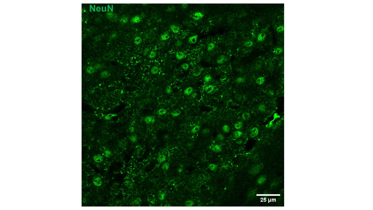
NeuN, encoded by FOX3, is a neuron-specific nuclear protein. Anti-NeuN stains exclusively neuronal cells in the central and peripheral nervous systems, especially postmitotic and differentiating neurons, as well as terminally differentiated neurons. Anti-NeuN has been used widely as a reliable tool to detect most postmitotic neuronal cell types. The immunohistochemical staining is primarily localized in the nucleus of the neurons with lighter staining in the cytoplasm.
Protocole
| Product Specific Protocols | |
|---|---|
| IHC protocol for NeuN antibody 66836-1-Ig | Download protocol |
| IF protocol for NeuN antibody 66836-1-Ig | Download protocol |
| Standard Protocols | |
|---|---|
| Click here to view our Standard Protocols |
Publications
| Species | Application | Title |
|---|---|---|
Cell Metab Acetate enables metabolic fitness and cognitive performance during sleep disruption | ||
Microbiome The microbiota-gut-brain axis participates in chronic cerebral hypoperfusion by disrupting the metabolism of short-chain fatty acids. | ||
Redox Biol LOX-mediated ECM mechanical stress induces Piezo1 activation in hypoxic-ischemic brain damage and identification of novel inhibitor of LOX | ||
Cell Death Dis ChemR23 activation attenuates cognitive impairment in chronic cerebral hypoperfusion by inhibiting NLRP3 inflammasome-induced neuronal pyroptosis | ||
Cell Death Dis Astrocyte-derived exosomal nicotinamide phosphoribosyltransferase (Nampt) ameliorates ischemic stroke injury by targeting AMPK/mTOR signaling to induce autophagy | ||
Diabetes Regulatory Role of NF-κB on HDAC2 and Tau Hyperphosphorylation in Diabetic Encephalopathy and the Therapeutic Potential of Luteolin |
Avis
The reviews below have been submitted by verified Proteintech customers who received an incentive for providing their feedback.
FH Deng (Verified Customer) (08-14-2025) | it works, but has some background (see image in mouse PVN region)
 |
FH Carla (Verified Customer) (02-03-2025) | We didn't have any good results
|
FH Kenzo (Verified Customer) (01-05-2024) | This monoclonal NeuN worked well for mouse tissues.
|
FH Silvia (Verified Customer) (08-11-2022) | it works well for Immunofluorescence on mature neurons
|
FH Delphine (Verified Customer) (07-25-2022) | Immunohistochemistry with the NeuN antibody on frozen spinal cord worked but the labeling must be optimized because there is a lot of background noise.
|
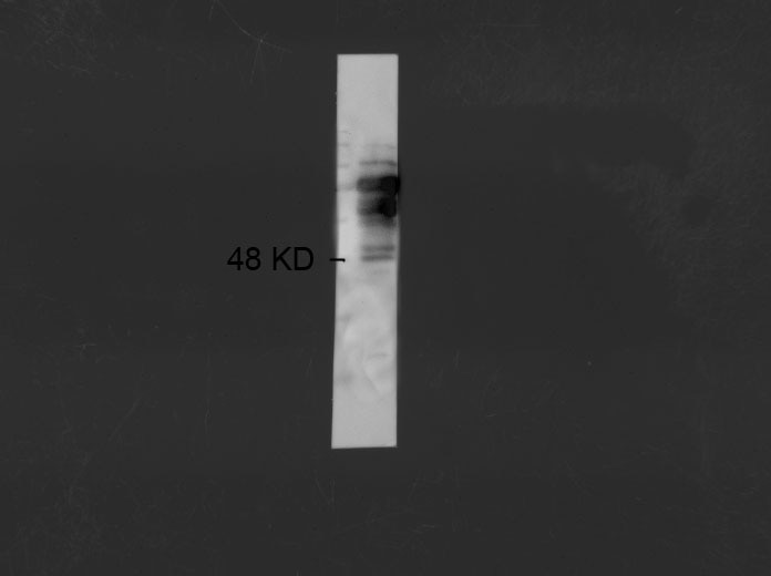
FH q (Verified Customer) (01-05-2022) | It is OK to use it in WB, but several non-specific bands above the expected MW.
 |

