- Phare
- Validé par KD/KO
Anticorps Polyclonal de lapin anti-OPA1
OPA1 Polyclonal Antibody for WB, IHC, IP, ELISA
Hôte / Isotype
Lapin / IgG
Réactivité testée
Humain, rat, souris et plus (7)
Applications
WB, IHC, IF, IP, CoIP, ELISA
Conjugaison
Non conjugué
N° de cat : 27733-1-AP
Synonymes
Galerie de données de validation
 at dilution of 1:15000 incubated at room temperature for 1.5 hours.")
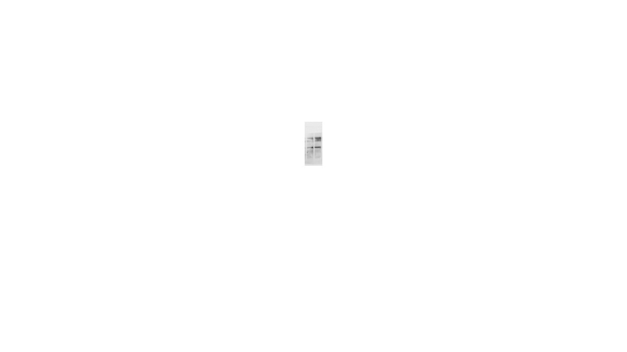
 with sh-Control and sh-OPA1 transfected HEK-293 cells.")
 at dilution of 1:2000 incubated at room temperature for 1.5 hours.")
 at dilution of 1:8000 incubated at room temperature for 1.5 hours.")
 at dilution of 1:6000 incubated at room temperature for 1.5 hours.")
 with mouse brain tissue lysate 1280 ug.")
 at dilution of 1:200 (under 10x lens. Heat mediated antigen retrieval with Tris-EDTA buffer (pH 9.0).")
 at dilution of 1:200 (under 40x lens. Heat mediated antigen retrieval with Tris-EDTA buffer (pH 9.0).")
 at dilution of 1:1000 (under 10x lens). Heat mediated antigen retrieval with Tris-EDTA buffer (pH 9.0).")
 at dilution of 1:1000 (under 40x lens). Heat mediated antigen retrieval with Tris-EDTA buffer (pH 9.0).")
 at dilution of 1:200 (under 40x lens). Heat mediated antigen retrieval with Tris-EDTA buffer (pH 9.0).")
 at dilution of 1:200 (under 10x lens. Heat mediated antigen retrieval with Tris-EDTA buffer (pH 9.0).")
 at dilution of 1:200 (under 40x lens. Heat mediated antigen retrieval with Tris-EDTA buffer (pH 9.0).")
 at dilution of 1:200 (under 40x lens. Heat mediated antigen retrieval with Tris-EDTA buffer (pH 9.0).")
 at dilution of 1:200 (under 10x lens. Heat mediated antigen retrieval with Tris-EDTA buffer (pH 9.0).")
Applications testées
| Résultats positifs en WB | cellules A431, cellules HEK-293, cellules HEK-293T, cellules HeLa, cellules HepG2, cellules L02, tissu cérébral de rat, tissu cérébral de souris, tissu hépatique de souris |
| Résultats positifs en IP | tissu cérébral de souris, |
| Résultats positifs en IHC | tissu cardiaque de souris, tissu cérébral de souris il est suggéré de démasquer l'antigène avec un tampon de TE buffer pH 9.0; (*) À défaut, 'le démasquage de l'antigène peut être 'effectué avec un tampon citrate pH 6,0. |
Dilution recommandée
| Application | Dilution |
|---|---|
| Western Blot (WB) | WB : 1:5000-1:50000 |
| Immunoprécipitation (IP) | IP : 0.5-4.0 ug for 1.0-3.0 mg of total protein lysate |
| Immunohistochimie (IHC) | IHC : 1:50-1:500 |
| It is recommended that this reagent should be titrated in each testing system to obtain optimal results. | |
| Sample-dependent, check data in validation data gallery | |
Applications publiées
| KD/KO | See 4 publications below |
| WB | See 194 publications below |
| IHC | See 8 publications below |
| IF | See 12 publications below |
| IP | See 1 publications below |
| CoIP | See 1 publications below |
Informations sur le produit
27733-1-AP cible OPA1 dans les applications de WB, IHC, IF, IP, CoIP, ELISA et montre une réactivité avec des échantillons Humain, rat, souris
| Réactivité | Humain, rat, souris |
| Réactivité citée | rat, bovin, Chèvre, Humain, poisson-zèbre, porc, poulet, souris, Hamster, duck |
| Hôte / Isotype | Lapin / IgG |
| Clonalité | Polyclonal |
| Type | Anticorps |
| Immunogène | OPA1 Protéine recombinante Ag26887 |
| Nom complet | optic atrophy 1 (autosomal dominant) |
| Masse moléculaire calculée | 960 aa, 112 kDa |
| Poids moléculaire observé | 80-100 kDa |
| Numéro d’acquisition GenBank | BC075805 |
| Symbole du gène | OPA1 |
| Identification du gène (NCBI) | 4976 |
| Conjugaison | Non conjugué |
| Forme | Liquide |
| Méthode de purification | Purification par affinité contre l'antigène |
| Tampon de stockage | PBS with 0.02% sodium azide and 50% glycerol |
| Conditions de stockage | Stocker à -20°C. Stable pendant un an après l'expédition. L'aliquotage n'est pas nécessaire pour le stockage à -20oC Les 20ul contiennent 0,1% de BSA. |
Informations générales
OPA1 is a nuclear-encoded mitochondrial protein with similarity to dynamin-related GTPases. OPA1 localizes to the inner mitochondrial membrane and helps regulate mitochondrial stability and energy output. This protein also sequesters cytochrome c. OPA1 is associated with the inner membrane and protects cells from apoptosis by regulating inner membrane dynamics. Mutation of OPA1 causes the disease dominant optic atrophy, a degeneration of the retinal ganglion cells. OPA1 undergoes complex posttranscriptional regulation and posttranslational proteolysis. OPA1 is regulated by proteolytic cleavage, which degrades long OPA1 isoforms into short isoforms. The gene OPA1 can be cleaved into some chains with MW 100 kDa and 80-90 kDa.
Protocole
| Product Specific Protocols | |
|---|---|
| WB protocol for OPA1 antibody 27733-1-AP | Download protocol |
| IHC protocol for OPA1 antibody 27733-1-AP | Download protocol |
| IP protocol for OPA1 antibody 27733-1-AP | Download protocol |
| Standard Protocols | |
|---|---|
| Click here to view our Standard Protocols |
Publications
| Species | Application | Title |
|---|---|---|
Cell Res Mitochondria-localized cGAS suppresses ferroptosis to promote cancer progression | ||
ACS Cent Sci Macrophage Inactivation by Small Molecule Wedelolactone via Targeting sEH for the Treatment of LPS-Induced Acute Lung Injury | ||
Mol Cell β-hydroxybutyrate facilitates mitochondrial-derived vesicle biogenesis and improves mitochondrial functions | ||
Mol Cell Serine synthesis sustains macrophage IL-1β production via NAD+-dependent protein acetylation | ||
Mol Cell Filamentous GLS1 promotes ROS-induced apoptosis upon glutamine deprivation via insufficient asparagine synthesis.
| ||
Acta Pharm Sin B Honokiol alleviated neurodegeneration by reducing oxidative stress and improving mitochondrial function in mutant SOD1 cellular and mouse models of amyotrophic lateral sclerosis |
Avis
The reviews below have been submitted by verified Proteintech customers who received an incentive for providing their feedback.
FH Kahimbi (Verified Customer) (01-31-2025) | It was used as a primary antibody for a western blot and the bands were clearly visible. The antibody worked well.
|
FH P (Verified Customer) (09-23-2024) | excellent!
 |
FH Mi (Verified Customer) (02-21-2023) | Works well in human adipocyte cells, we got clean bands at the expected size.
|

