- Phare
- Validé par KD/KO
Anticorps Polyclonal de lapin anti-P53
P53 Polyclonal Antibody for WB, IHC, IF/ICC, IP, ELISA
Hôte / Isotype
Lapin / IgG
Réactivité testée
Humain, rat et plus (6)
Applications
WB, IHC, IF/ICC, IP, CoIP, ChIP, RIP, ELISA
Conjugaison
Non conjugué
N° de cat : 10442-1-AP
Synonymes
Galerie de données de validation
 at dilution of 1:3000 incubated at room temperature for 1.5 hours.")
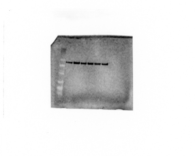
 with sh-Control and sh-P53 transfected A431 cells.")
 at dilution of 1:20000 incubated at room temperature for 1.5 hours.")
 at dilution of 1:10000 incubated at room temperature for 1.5 hours.")
 at dilution of 1:1000 incubated at room temperature for 1.5 hours.")
 at dilution of 1:300 incubated at room temperature for 1.5 hours.")
 at dilution of 1:3000 incubated at room temperature for 1.5 hours.")
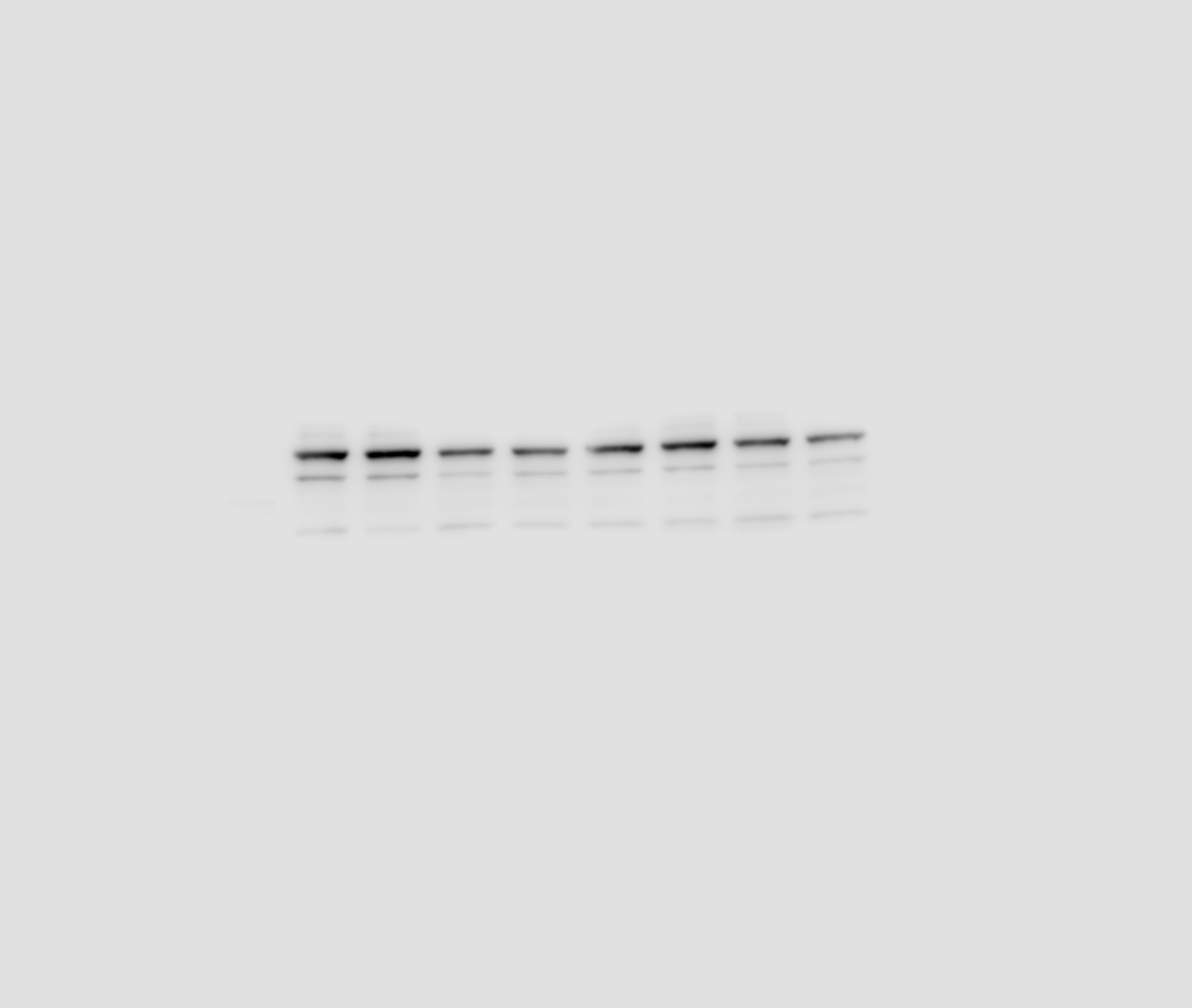
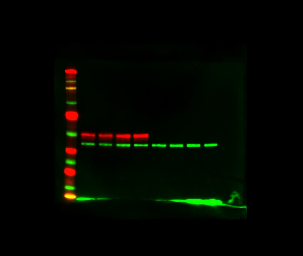
 with HEK-293 cells lysate 1560 ug.")
 at dilution of 1:500 (under 20x lens). Heat mediated antigen retrieval with Tris-EDTA buffer (pH 9.0).")
 at dilution of 1:500 (under 20x lens). Heat mediated antigen retrieval with Tris-EDTA buffer (pH 9.0).")
 fixed A431 cells using P53 antibody (10442-1-AP) at dilution of 1:400 and CoraLite®488-Conjugated AffiniPure Goat Anti-Rabbit IgG(H+L), CL594-Phalloidin (red).")
 fixed A431 cells using P53 antibody (10442-1-AP) at dilution of 1:400 and CoraLite®488-Conjugated AffiniPure Goat Anti-Rabbit IgG(H+L).")
Applications testées
| Résultats positifs en WB | cellules A431, cellules HEK-293, cellules HepG2, cellules HT-29, cellules MCF-7, cellules SMMC-7721, tissu de côlon de rat |
| Résultats positifs en IP | cellules HEK-293, |
| Résultats positifs en IHC | human ovary cancer tissue, il est suggéré de démasquer l'antigène avec un tampon de TE buffer pH 9.0; (*) À défaut, 'le démasquage de l'antigène peut être 'effectué avec un tampon citrate pH 6,0. |
| Résultats positifs en IF/ICC | cellules A431, |
Dilution recommandée
| Application | Dilution |
|---|---|
| Western Blot (WB) | WB : 1:5000-1:50000 |
| Immunoprécipitation (IP) | IP : 0.5-4.0 ug for 1.0-3.0 mg of total protein lysate |
| Immunohistochimie (IHC) | IHC : 1:250-1:1000 |
| Immunofluorescence (IF)/ICC | IF/ICC : 1:200-1:800 |
| It is recommended that this reagent should be titrated in each testing system to obtain optimal results. | |
| Sample-dependent, check data in validation data gallery | |
Informations sur le produit
10442-1-AP cible P53 dans les applications de WB, IHC, IF/ICC, IP, CoIP, ChIP, RIP, ELISA et montre une réactivité avec des échantillons Humain, rat
| Réactivité | Humain, rat |
| Réactivité citée | rat, Chèvre, Humain, Lapin, poisson-zèbre, porc, poulet, singe |
| Hôte / Isotype | Lapin / IgG |
| Clonalité | Polyclonal |
| Type | Anticorps |
| Immunogène | P53 Protéine recombinante Ag0698 |
| Nom complet | tumor protein p53 |
| Masse moléculaire calculée | 44 kDa |
| Poids moléculaire observé | 53 kDa |
| Numéro d’acquisition GenBank | BC003596 |
| Symbole du gène | P53 |
| Identification du gène (NCBI) | 7157 |
| Conjugaison | Non conjugué |
| Forme | Liquide |
| Méthode de purification | Purification par affinité contre l'antigène |
| Tampon de stockage | PBS with 0.02% sodium azide and 50% glycerol |
| Conditions de stockage | Stocker à -20°C. Stable pendant un an après l'expédition. L'aliquotage n'est pas nécessaire pour le stockage à -20oC Les 20ul contiennent 0,1% de BSA. |
Informations générales
1. What is p53?
P53 is a tumor suppressor gene that plays a role in maintaining genomic stability and controlling apoptosis. During the cell cycle, it can arrest cells at the G1/S checkpoint and activate DNA repair mechanisms. It is the most mutated gene in cancer. In unstressed cells, p53 usually exists at low levels in an inactive form, being bound to Mdm2.
2. FAQs and p53
a. I fail to detect p53 by western blotting
Basal levels of wild-type p53 in untreated cells can be low. Try to load more cell lysate and use a positive control - a lysate of cells treated with DNA-damaging agents should increase p53 levels.
b. I fail to detect p53 in some cell lines by western blotting
Various p53 mutations are present in cancer cell types. If mutations cause truncations/deletions some monoclonal antibodies may no longer recognize mutated p53. You have more chances of detecting various p53 mutants with our polyclonal antibody.
c. I can detect more than one band ~50 kDa size / different cell lines give bands at slightly different size
p53 is a subject of post-translational modifications (http://p53.free.fr/p53_info/p53_modifications.html) and more than one isoform may be expressed (http://p53.free.fr/p53_info/p53_isoforms.html). Also, it is possible that your cell line of interest expresses one allele with mutated p53 with altered molecular weight.
Protocole
| Product Specific Protocols | |
|---|---|
| WB protocol for P53 antibody 10442-1-AP | Download protocol |
| IHC protocol for P53 antibody 10442-1-AP | Download protocol |
| IF protocol for P53 antibody 10442-1-AP | Download protocol |
| IP protocol for P53 antibody 10442-1-AP | Download protocol |
| Standard Protocols | |
|---|---|
| Click here to view our Standard Protocols |
Publications
| Species | Application | Title |
|---|---|---|
ACS Nano Mesenchymal Stem Cell-Derived Extracellular Vesicles Attenuate Mitochondrial Damage and Inflammation by Stabilizing Mitochondrial DNA. | ||
Nat Commun URI alleviates tyrosine kinase inhibitors-induced ferroptosis by reprogramming lipid metabolism in p53 wild-type liver cancers | ||
Nat Commun High-content screening identifies ganoderic acid A as a senotherapeutic to prevent cellular senescence and extend healthspan in preclinical models | ||
Sci Transl Med PTEN status determines chemosensitivity to proteasome inhibition in cholangiocarcinoma. | ||
Cell Res DNA damage triggers tubular endoplasmic reticulum extension to promote apoptosis by facilitating ER-mitochondria signaling. |
Avis
The reviews below have been submitted by verified Proteintech customers who received an incentive for providing their feedback.
FH Courtney (Verified Customer) (08-28-2025) | Bands turned out great with no background. I didn't have to do any optimizing.
 |
FH Angelique (Verified Customer) (06-26-2025) | very nice expression
|
FH Laetitia (Verified Customer) (03-21-2024) | Finding antibodies specific to avian proteins is very difficult. I have tried different antibodies from different companies so far. This antibody is by far the best (cost efficient as well)
|
FH Macarena Lucia (Verified Customer) (10-17-2022) | clear band on human lysates
|
FH Kimberly (Verified Customer) (04-08-2022) | TRIS antigen retrieval
|
FH Ryan (Verified Customer) (08-27-2021) | Worked well in HeLa cells and provided a nice clear band.
|
FH Fan (Verified Customer) (03-22-2021) | Mouse retinal tissue was used. Single band at the right size was detected. Good antibody.
 |
FH WEI (Verified Customer) (01-17-2020) | Weak band in heart lysates
|
FH Ben (Verified Customer) (09-26-2019) | It worked very well for our westerns.
|
FH Sid (Verified Customer) (04-12-2019) | Product performed well in western for human samples, moderate background/non specific binding
|
FH Josiah (Verified Customer) (02-27-2019) | Antibody performed superb in a Western Blot application. Used Licor IR secondary antibodies to label the p53 antibody along with ß-actin as a duplex assay and imaged on a Licor Odyssey FC.
 |

