- Phare
- Validé par KD/KO
Anticorps Polyclonal de lapin anti-PARP9
PARP9 Polyclonal Antibody for WB, IHC, IP, ELISA
Hôte / Isotype
Lapin / IgG
Réactivité testée
Humain, rat
Applications
WB, IHC, IF, IP, ELISA
Conjugaison
Non conjugué
N° de cat : 17535-1-AP
Synonymes
Galerie de données de validation
Applications testées
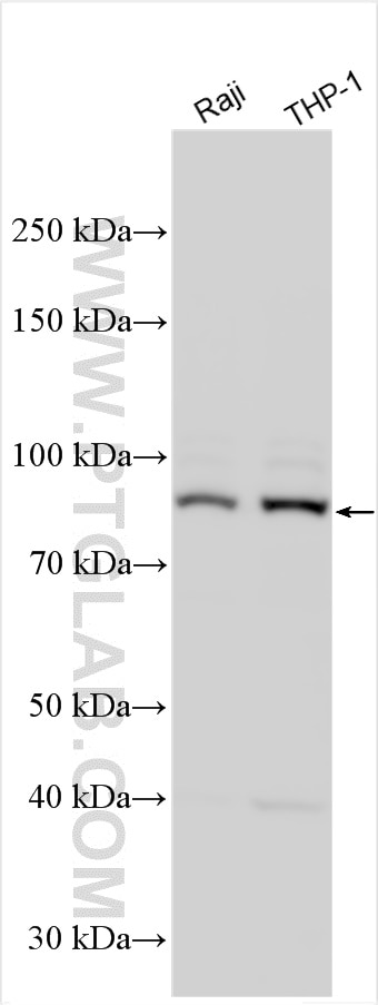
| Résultats positifs en WB | cellules Raji, cellules THP-1 |
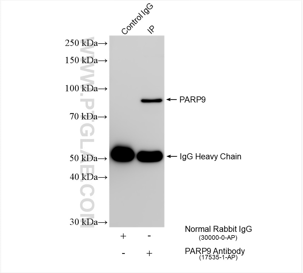
| Résultats positifs en IP | cellules MCF-7, |
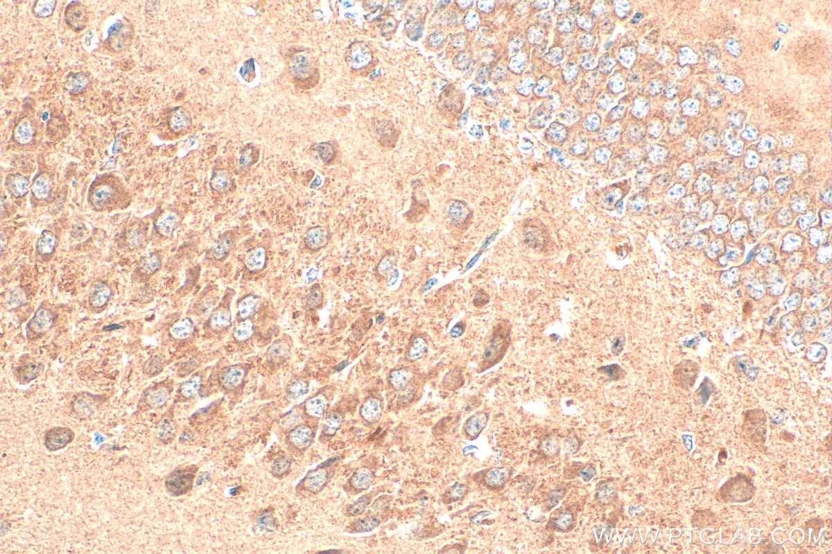
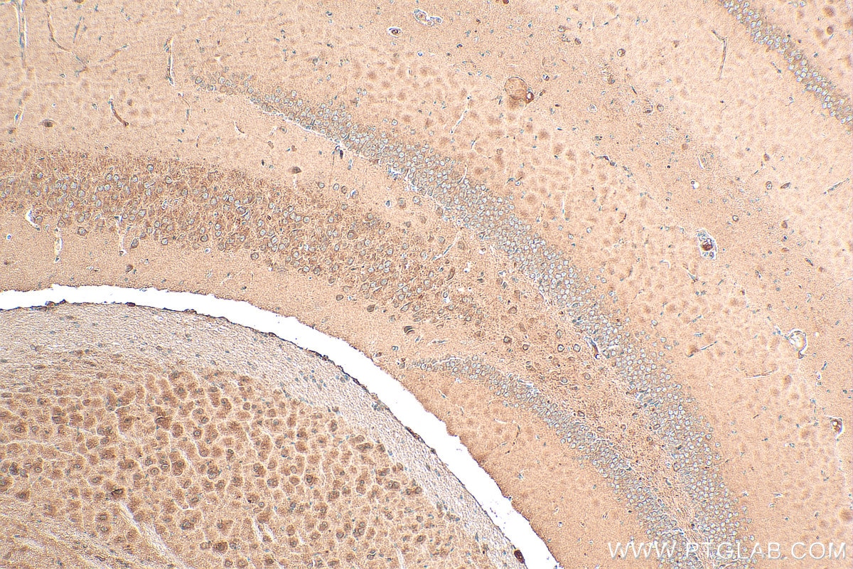
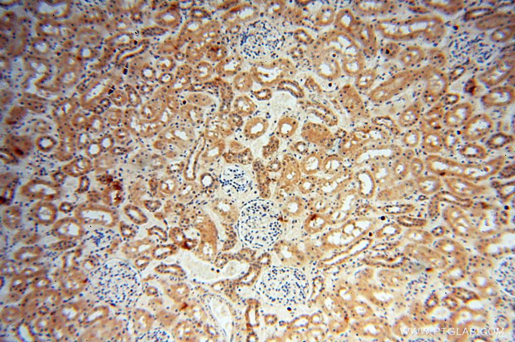
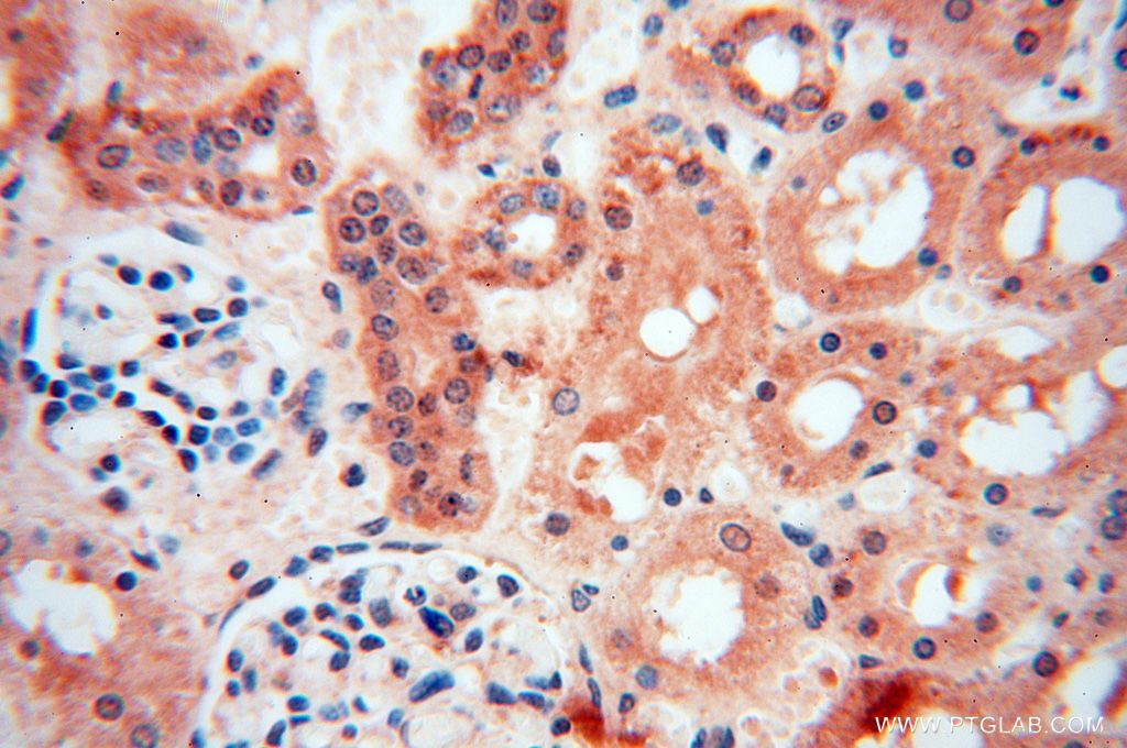
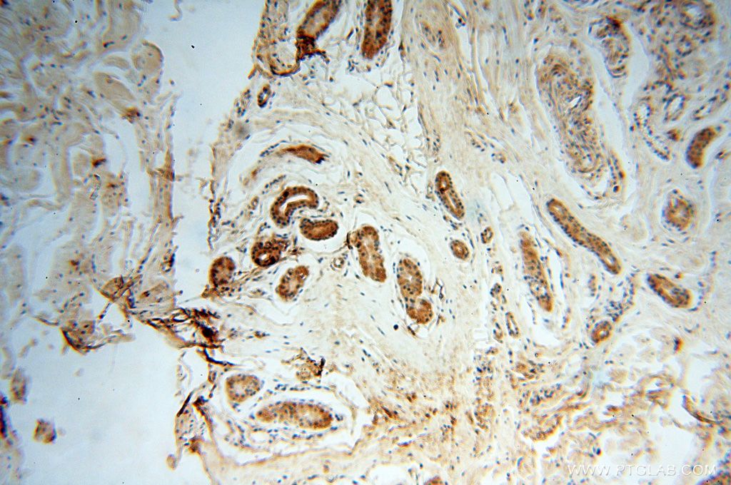
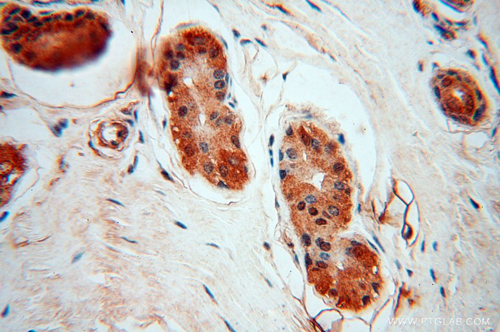
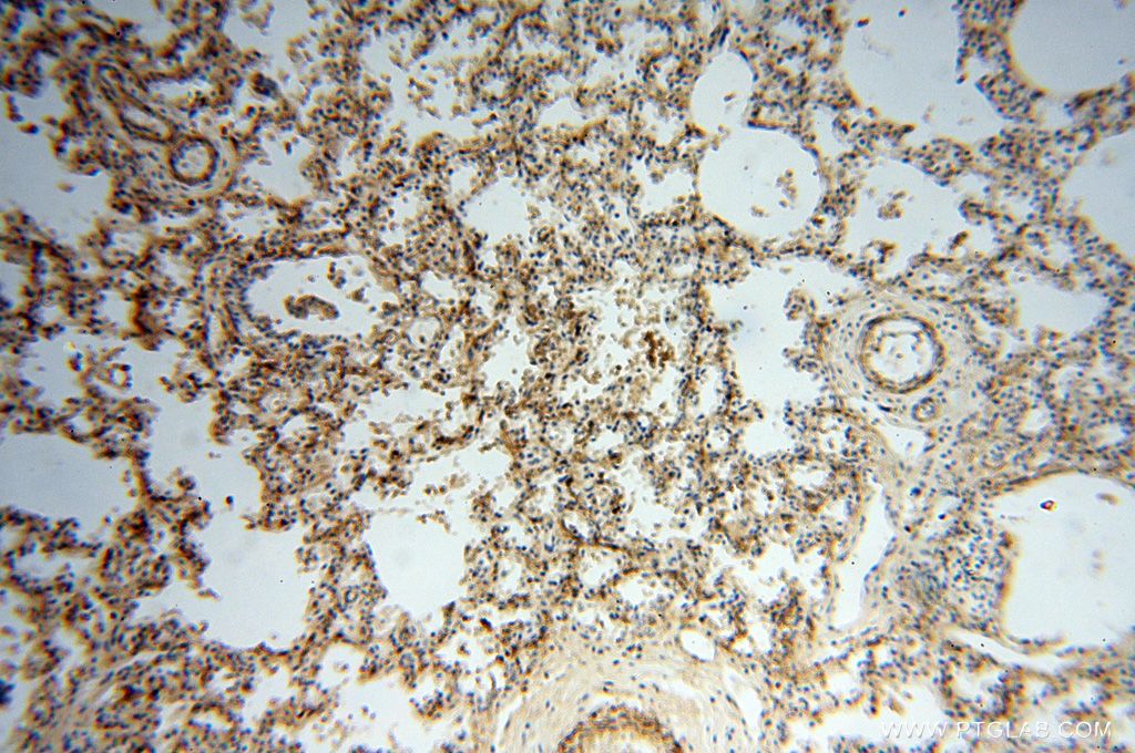
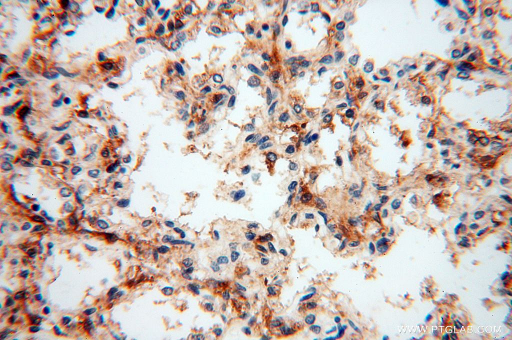
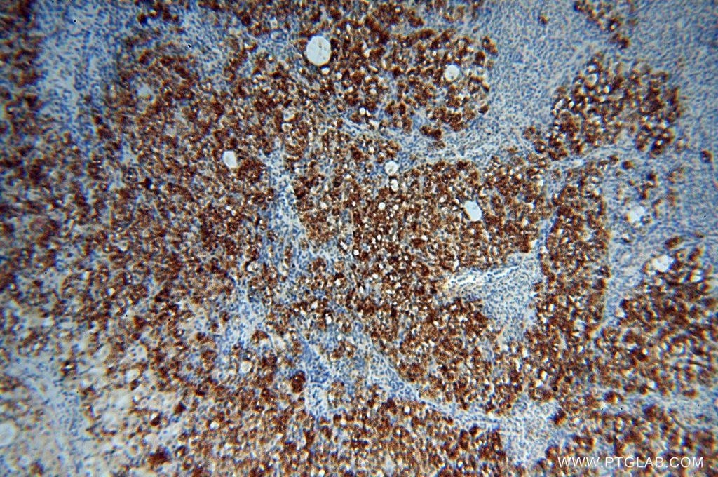
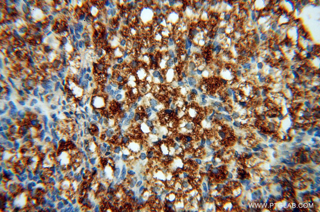
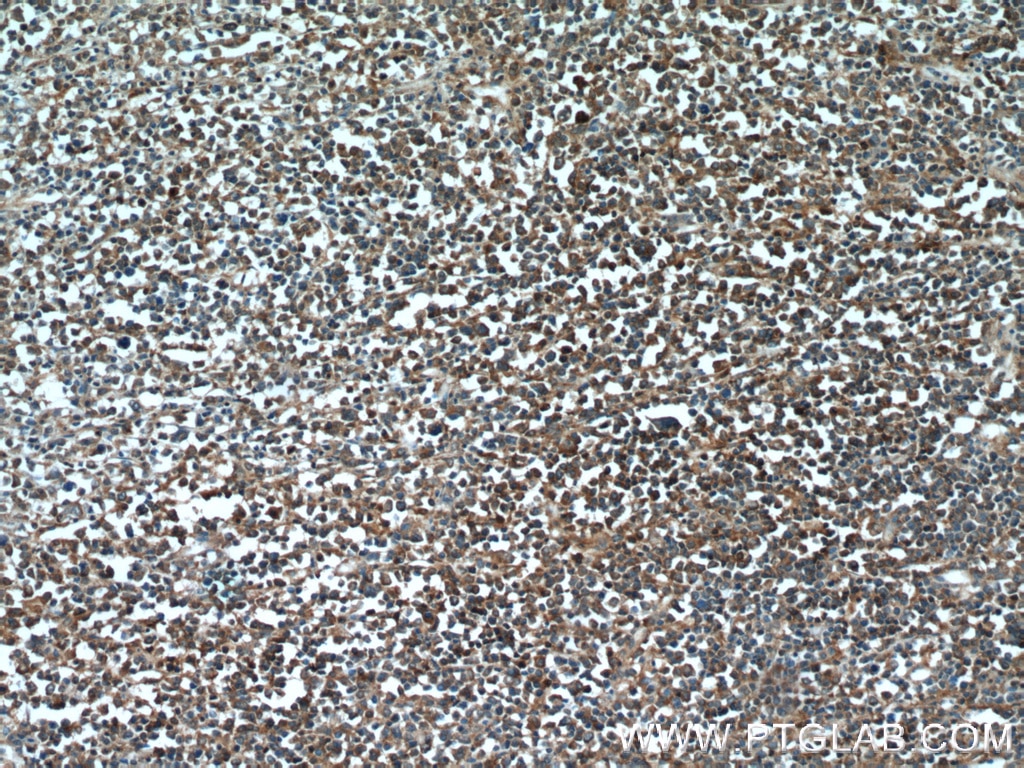
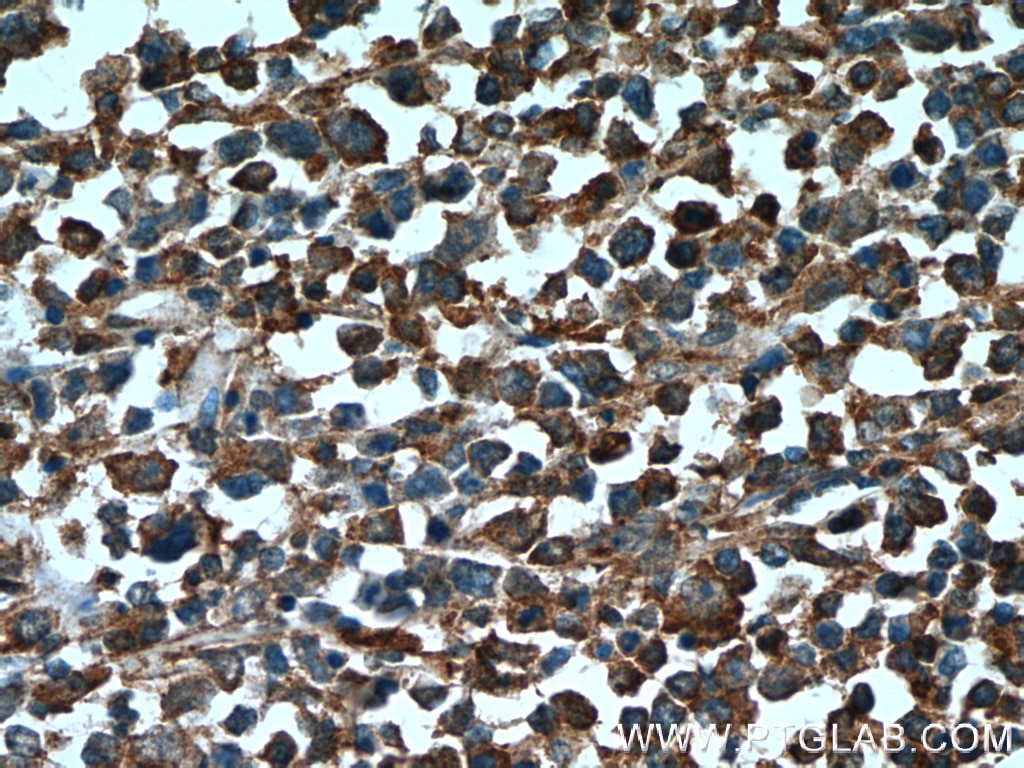
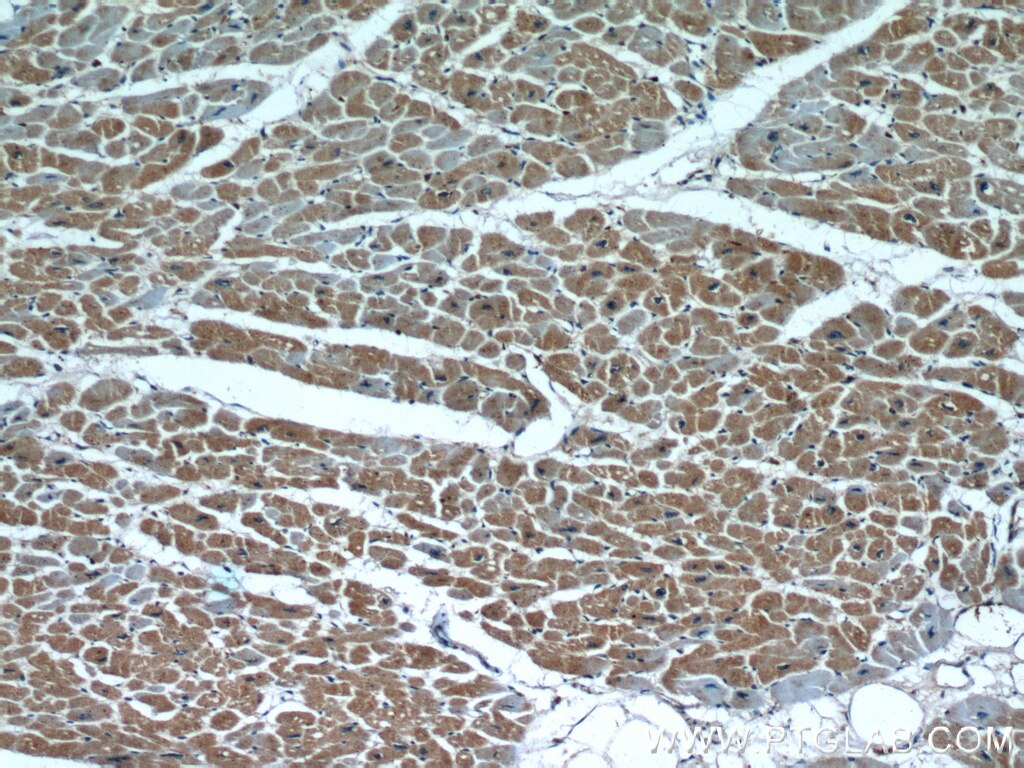
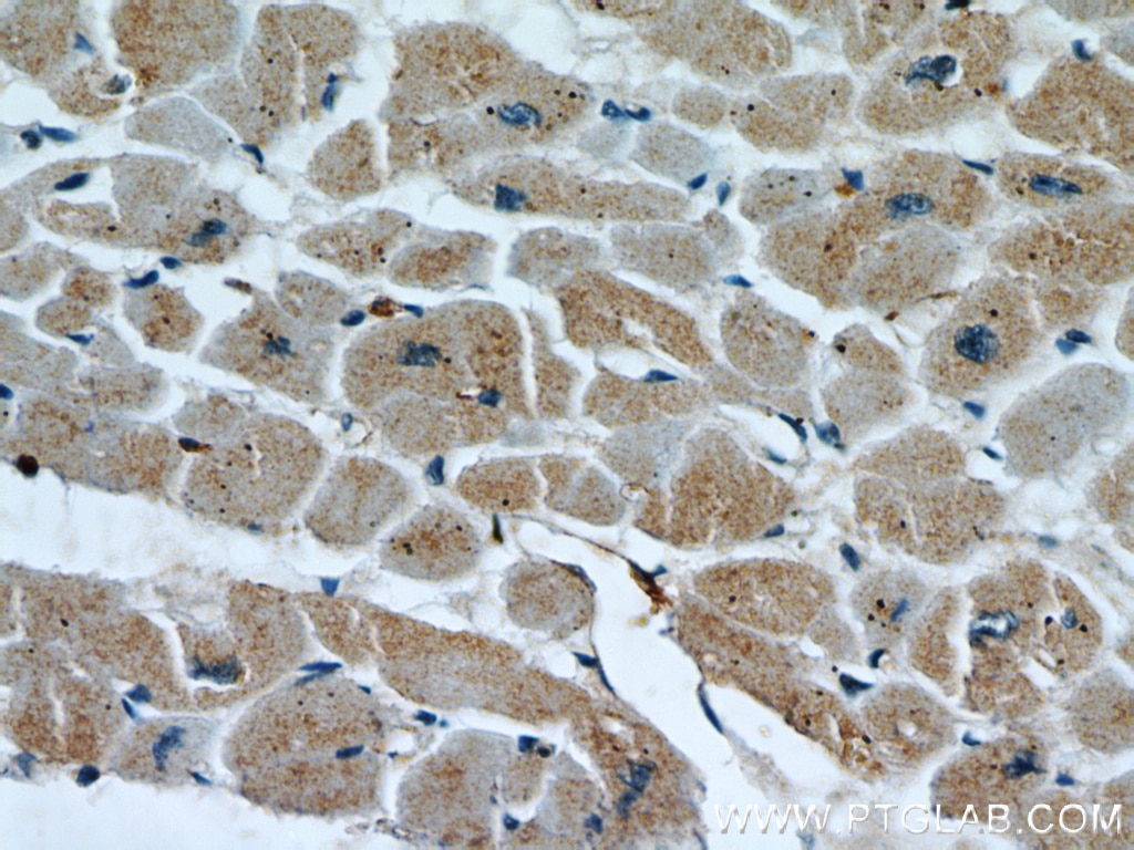
| Résultats positifs en IHC | tissu cérébral de souris, tissu cardiaque humain, tissu cutané humain, tissu de lymphome humain, tissu ovarien humain, tissu pulmonaire humain, tissu rénal humain il est suggéré de démasquer l'antigène avec un tampon de TE buffer pH 9.0; (*) À défaut, 'le démasquage de l'antigène peut être 'effectué avec un tampon citrate pH 6,0. |
Dilution recommandée
| Application | Dilution |
|---|---|
| Western Blot (WB) | WB : 1:2000-1:12000 |
| Immunoprécipitation (IP) | IP : 0.5-4.0 ug for 1.0-3.0 mg of total protein lysate |
| Immunohistochimie (IHC) | IHC : 1:250-1:1000 |
| It is recommended that this reagent should be titrated in each testing system to obtain optimal results. | |
| Sample-dependent, check data in validation data gallery | |
Applications publiées
| KD/KO | See 4 publications below |
| WB | See 6 publications below |
| IHC | See 1 publications below |
| IF | See 2 publications below |
Informations sur le produit
17535-1-AP cible PARP9 dans les applications de WB, IHC, IF, IP, ELISA et montre une réactivité avec des échantillons Humain, rat
| Réactivité | Humain, rat |
| Réactivité citée | Humain |
| Hôte / Isotype | Lapin / IgG |
| Clonalité | Polyclonal |
| Type | Anticorps |
| Immunogène | PARP9 Protéine recombinante Ag11587 |
| Nom complet | poly (ADP-ribose) polymerase family, member 9 |
| Masse moléculaire calculée | 819 aa, 92 kDa |
| Poids moléculaire observé | 88 kDa |
| Numéro d’acquisition GenBank | BC039580 |
| Symbole du gène | PARP9 |
| Identification du gène (NCBI) | 83666 |
| Conjugaison | Non conjugué |
| Forme | Liquide |
| Méthode de purification | Purification par affinité contre l'antigène |
| Tampon de stockage | PBS with 0.02% sodium azide and 50% glycerol |
| Conditions de stockage | Stocker à -20°C. Stable pendant un an après l'expédition. L'aliquotage n'est pas nécessaire pour le stockage à -20oC Les 20ul contiennent 0,1% de BSA. |
Informations générales
Poly(ADP-ribosyl)ation is a post-translational modification of proteins mediated by one of the 17 members of the poly(ADP-ribose) polymerases (PARP). PARP-9 belongs to the subfamily of macroPARPs, associating one to three macro domains to the PARP domain. Overexpression of PARP-9 stimulates cell migration in vitro, suggesting a role for PARP-9 in the promotion of malignant B cell migration and dissemination in high risk DLBCL. PARP-9 is also likely a transcription coactivator, its overexpression in B lymphocytes, stimulated by IFNγ, inducing the transcription of IFNγ-controlled genes.
Protocole
| Product Specific Protocols | |
|---|---|
| WB protocol for PARP9 antibody 17535-1-AP | Download protocol |
| IHC protocol for PARP9 antibody 17535-1-AP | Download protocol |
| IP protocol for PARP9 antibody 17535-1-AP | Download protocol |
| Standard Protocols | |
|---|---|
| Click here to view our Standard Protocols |
Publications
| Species | Application | Title |
|---|---|---|
J Immunol An Alternative Splicing of Tupaia STING Modulated Anti-RNA Virus Responses by Targeting MDA5-LGP2 and IRF3.
| ||
J Biol Chem Patient-derived organotypic tissue cultures as a platform to evaluate metabolic reprogramming in breast cancer patients | ||
Oncol Lett PARP9 is overexpressed in human breast cancer and promotes cancer cell migration.
| ||
J Mol Biol KH-like domains in PARP9/DTX3L and PARP14 coordinate protein-protein interactions to promote cancer cell survival
|
Avis
The reviews below have been submitted by verified Proteintech customers who received an incentive for providing their feedback.
FH Lisa (Verified Customer) (07-23-2025) | Good working antibody for WB.
|
FH Hadil (Verified Customer) (05-25-2023) | I have had trouble detecting PARP9 signals with other PARP9 antibodies, however, this antibody is a game changer. It works well both at room temperature (1.5 -2 hrs) and also overnight at 1:500 dilution. Would highly recommend to anyone who's studying PARP9.
|
