- Phare
- Validé par KD/KO
Anticorps Monoclonal anti-PD-1/CD279
PD-1/CD279 Monoclonal Antibody for WB, IHC, IF-P, ELISA
Hôte / Isotype
Mouse / IgG2b
Réactivité testée
Humain, porc, rat, souris
Applications
WB, IHC, IF-P, IP, ELISA
Conjugaison
Non conjugué
CloneNo.
4H4D1
N° de cat : 66220-1-Ig
Synonymes
Galerie de données de validation
 at dilution of 1:15000 incubated at room temperature for 1.5 hours. The membrane was stripped and reblotted with HRP-conjugated Lamin B1 Monoclonal antibody (HRP-66095) as loading control.")
 at dilution of 1:15000 incubated at room temperature for 1.5 hours. The membrane was stripped and reblotted with HRP-conjugated Lamin B1 Monoclonal antibody (HRP-66095) as loading control.")
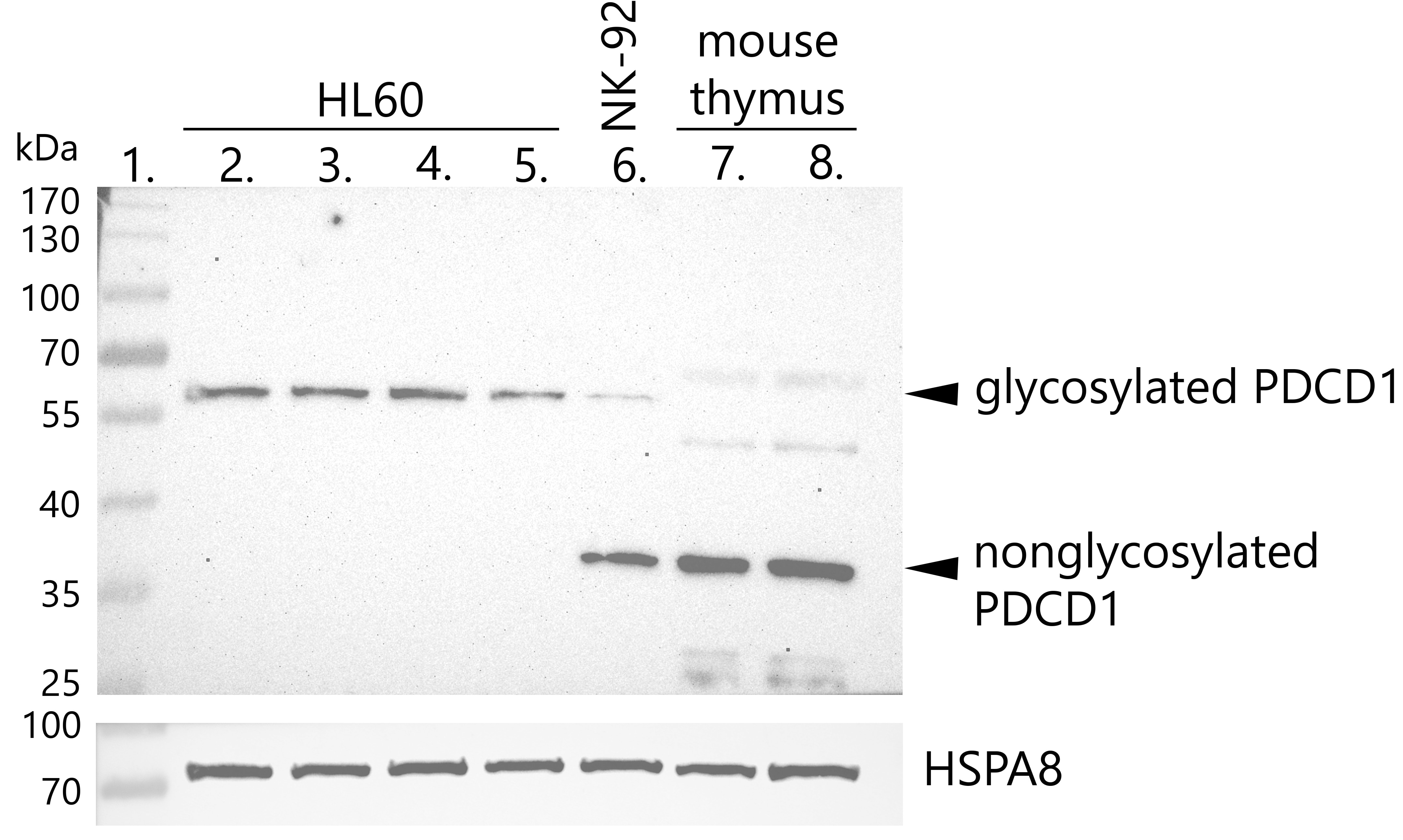
 at dilution of 1:5000 incubated at room temperature for 1.5 hours.")
 at dilution of 1:1000 incubated at room temperature for 1.5 hours.")
 at dilution of 1:5000 incubated at room temperature for 1.5 hours.")
 at dilution of 1:4000 (under 20x lens). Heat mediated antigen retrieval with Tris-EDTA buffer (pH 9.0).")
 at dilution of 1:20000 (under 10x lens). Heat mediated antigen retrieval with Tris-EDTA buffer (pH 9.0).")
 at dilution of 1:10000 (under 10x lens). Heat mediated antigen retrieval with Tris-EDTA buffer (pH 9.0).")
 at dilution of 1:10000 (under 40x lens). Heat mediated antigen retrieval with Tris-EDTA buffer (pH 9.0).")
 at dilution of 1:200 (under 10x lens). . Heat mediated antigen retrieved with Tris-EDTA buffer, pH9.0.")
 at dilution of 1:200 (under 40x lens). . Heat mediated antigen retrieved with Tris-EDTA buffer, pH9.0.")
 fixed human tonsillitis tissue using PD-1/CD279 mouse mAb (66220-1-Ig) at dilution of 1:50 and CD20 rabbit pAb (24828-1-AP) at dilution of 1:50, further stained with Alexa Fluor 488-conjugated AffiniPure Goat Anti-Mouse IgG(H+L) for 66220-1-Ig, and Alexa Fluor 594-conjugated AffiniPure Goat Anti-Rabbit IgG (H+L) for 24828-1-AP.")
 fixed paraffin-embedded human angioimmunoblastic T-cell lymphoma tissue using PD-1/CD279 antibody (66220-1-Ig, Clone: 4H4D1 ) at dilution of 1:400 and CoraLite®488-Conjugated AffiniPure Goat Anti-Mouse IgG(H+L) (SA00013-1). Heat mediated antigen retrieval with Tris-EDTA buffer (pH 9.0).")
 fixed paraffin-embedded human angioimmunoblastic T-cell lymphoma tissue using PD-1/CD279 antibody (66220-1-Ig, Clone: 4H4D1 ) at dilution of 1:400 and CoraLite®488-Conjugated AffiniPure Goat Anti-Mouse IgG(H+L) (SA00013-1). Heat mediated antigen retrieval with Tris-EDTA buffer (pH 9.0).")
 fixed paraffin-embedded human lymphoma tissue using PD-1/CD279 antibody (66220-1-Ig, Clone: 4H4D1 ) at dilution of 1:400 and Multi-rAb CoraLite ® Plus 488-Goat Anti-Mouse Recombinant Secondary Antibody (H+L) (RGAM002). Heat mediated antigen retrieval with Tris-EDTA buffer (pH 9.0).")
 fixed paraffin-embedded human lymphoma tissue using PD-1/CD279 antibody (66220-1-Ig, Clone: 4H4D1 ) at dilution of 1:400 and Multi-rAb CoraLite ® Plus 488-Goat Anti-Mouse Recombinant Secondary Antibody (H+L) (RGAM002). Heat mediated antigen retrieval with Tris-EDTA buffer (pH 9.0).")
 fixed paraffin-embedded human tonsillitis tissue using PD-1/CD279 antibody (66220-1-Ig, Clone: 4H4D1 ) at dilution of 1:400 and CoraLite®488-Conjugated Goat Anti-Mouse IgG(H+L) (SA00013-1). Heat mediated antigen retrieval with Tris-EDTA buffer (pH 9.0).")
 fixed human tonsillitis tissue using PD-1/CD279 mouse mAb (66220-1-Ig) at dilution of 1:50 and CD20 rabbit pAb (24828-1-AP) at dilution of 1:50, further stained with Alexa Fluor 488-conjugated AffiniPure Goat Anti-Mouse IgG(H+L) for 66220-1-Ig, and Alexa Fluor 594-conjugated AffiniPure Goat Anti-Rabbit IgG (H+L) for 24828-1-AP.")
Applications testées
| Résultats positifs en WB | cellules RAW 264.7, cellules Jurkat, cellules MOLT-4, cellules THP-1, tissu de ganglion lymphatique humain, tissu de thymus de porc, tissu de thymus de souris, tissu splénique de rat |
| Résultats positifs en IHC | tissu d'amygdalite humain, tissu de lymphome humain il est suggéré de démasquer l'antigène avec un tampon de TE buffer pH 9.0; (*) À défaut, 'le démasquage de l'antigène peut être 'effectué avec un tampon citrate pH 6,0. |
| Résultats positifs en IF-P | tissu d'amygdalite humain, tissu de lymphome humain |
Dilution recommandée
| Application | Dilution |
|---|---|
| Western Blot (WB) | WB : 1:5000-1:50000 |
| Immunohistochimie (IHC) | IHC : 1:2000-1:8000 |
| Immunofluorescence (IF)-P | IF-P : 1:200-1:800 |
| It is recommended that this reagent should be titrated in each testing system to obtain optimal results. | |
| Sample-dependent, check data in validation data gallery | |
Applications publiées
| KD/KO | See 2 publications below |
| WB | See 37 publications below |
| IHC | See 19 publications below |
| IF | See 28 publications below |
| IP | See 1 publications below |
Informations sur le produit
66220-1-Ig cible PD-1/CD279 dans les applications de WB, IHC, IF-P, IP, ELISA et montre une réactivité avec des échantillons Humain, porc, rat, souris
| Réactivité | Humain, porc, rat, souris |
| Réactivité citée | rat, Humain, souris |
| Hôte / Isotype | Mouse / IgG2b |
| Clonalité | Monoclonal |
| Type | Anticorps |
| Immunogène | PD-1/CD279 Protéine recombinante Ag12470 |
| Nom complet | programmed cell death 1 |
| Masse moléculaire calculée | 288 aa, 32 kDa |
| Poids moléculaire observé | 32 kDa, 47-55 kDa |
| Numéro d’acquisition GenBank | BC074740 |
| Symbole du gène | PD-1 |
| Identification du gène (NCBI) | 5133 |
| Conjugaison | Non conjugué |
| Forme | Liquide |
| Méthode de purification | Purification par protéine A |
| Tampon de stockage | PBS with 0.02% sodium azide and 50% glycerol |
| Conditions de stockage | Stocker à -20°C. Stable pendant un an après l'expédition. L'aliquotage n'est pas nécessaire pour le stockage à -20oC Les 20ul contiennent 0,1% de BSA. |
Informations générales
Programmed cell death 1 (PD-1, also known as CD279) is an immunoinhibitory receptor that belongs to the CD28/CTLA-4 subfamily of the Ig superfamily. It is a 288 amino acid (aa) type I transmembrane protein composed of one Ig superfamily domain, a stalk, a transmembrane domain, and an intracellular domain containing an immunoreceptor tyrosine-based inhibitory motif (ITIM) as well as an immunoreceptor tyrosine-based switch motif (ITSM) (PMID: 18173375). PD-1 is expressed during thymic development and is induced in a variety of hematopoietic cells in the periphery by antigen receptor signaling and cytokines (PMID: 20636820). Engagement of PD-1 by its ligands PD-L1 or PD-L2 transduces a signal that inhibits T-cell proliferation, cytokine production, and cytolytic function (PMID: 19426218). It is critical for the regulation of T cell function during immunity and tolerance. Blockade of PD-1 can overcome immune resistance and also has been shown to have antitumor activity (PMID: 22658127; 23169436). The calculated molecular weight of PD-1 is 32 kDa. It has been reported that PD-1 is heavily glycosylated and migrates with an apparent molecular mass of 47-55 kDa on SDS-PAGE (PMID: 8671665; 17640856; 17003438).
Protocole
| Product Specific Protocols | |
|---|---|
| WB protocol for PD-1/CD279 antibody 66220-1-Ig | Download protocol |
| IHC protocol for PD-1/CD279 antibody 66220-1-Ig | Download protocol |
| IF protocol for PD-1/CD279 antibody 66220-1-Ig | Download protocol |
| Standard Protocols | |
|---|---|
| Click here to view our Standard Protocols |
Publications
| Species | Application | Title |
|---|---|---|
Nat Commun ERK and USP5 govern PD-1 homeostasis via deubiquitination to modulate tumor immunotherapy | ||
Adv Sci (Weinh) PPY-Induced iCAFs Cultivate an Immunosuppressive Microenvironment in Pancreatic Cancer | ||
Nat Commun m6A mRNA demethylase FTO regulates melanoma tumorigenicity and response to anti-PD-1 blockade. | ||
J Exp Clin Cancer Res BAP1 regulates HSF1 activity and cancer immunity in pancreatic cancer | ||
J Transl Med UBE2J1 is identified as a novel plasma cell-related gene involved in the prognosis of high-grade serous ovarian cancer | ||
Cancers (Basel) Overcoming PD-1 Inhibitor Resistance with a Monoclonal Antibody to Secreted Frizzled-Related Protein 2 in Metastatic Osteosarcoma. |
Avis
The reviews below have been submitted by verified Proteintech customers who received an incentive for providing their feedback.
FH Wiesława (Verified Customer) (12-08-2022) | works very well
 |
FH Macarena Lucia (Verified Customer) (10-17-2022) | clear band
|
FH Mona (Verified Customer) (10-17-2021) | Staining of paraffin-embedded lung tissue from hamster. Antigen retrieval was performed with Tris-EDTA (pH 9)
|

