- Phare
- Validé par KD/KO
Anticorps Monoclonal anti-PD-L1/CD274
PD-L1/CD274 Monoclonal Antibody for WB, IHC, IF/ICC, IF-P, ELISA
Hôte / Isotype
Mouse / IgG1
Réactivité testée
Humain, porc, rat, souris
Applications
WB, IHC, IF/ICC, IF-P, IP, CoIP, ChIP, ELISA
Conjugaison
Non conjugué
CloneNo.
2B11D11
N° de cat : 66248-1-Ig
Synonymes
Galerie de données de validation
 at dilution of 1:4000 incubated at room temperature for 1.5 hours.")
 at dilution of 1:5000 incubated at room temperature for 1.5 hours.")
 at dilution of 1:1000 incubated at room temperature for 1.5 hours.")
 at dilution of 1:1000 incubated at room temperature for 1.5 hours.")
 at dilution of 1:1000 incubated at room temperature for 1.5 hours.")
 at dilution of 1:1000 incubated at room temperature for 1.5 hours.")
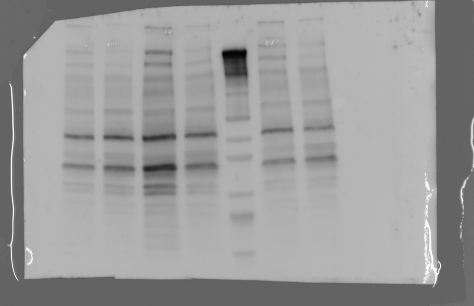
 with si-Control and si-PDL1 transfected HepG2 cells.")
 at dilution of 1:5000 incubated at room temperature for 1.5 hours.")
 at dilution of 1:5000 incubated at room temperature for 1.5 hours. PNGase F was obtained from Atagenix (cat.NO. ata808).")
 at dilution of 1:10000 (under 4x lens. Heat mediated antigen retrieval with Tris-EDTA buffer (pH 9.0).")
 at dilution of 1:10000 (under 10x lens. Heat mediated antigen retrieval with Tris-EDTA buffer (pH 9.0).")
 at dilution of 1:20000 (under 10x lens). Heat mediated antigen retrieval with Tris-EDTA buffer (pH 9.0). Slide was treated with PNGase F before staining.")
 at dilution of 1:20000 (under 40x lens). Heat mediated antigen retrieval with Tris-EDTA buffer (pH 9.0). Slide was treated with PNGase F before staining.")
 at dilution of 1:500 (under 10x lens).")
 at dilution of 1:500 (under 40x lens).")
 at dilution of 1:1000 (under 10x lens). Heat mediated antigen retrieval with Tris-EDTA buffer (pH 9.0).")
 at dilution of 1:1000 (under 40x lens). Heat mediated antigen retrieval with Tris-EDTA buffer (pH 9.0).")
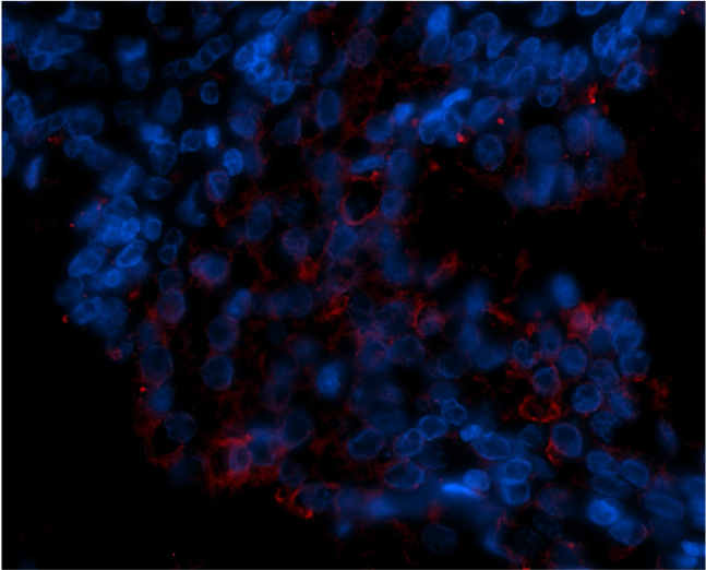
 fixed paraffin-embedded human placenta tissue using PD-L1/CD274 antibody (66248-1-Ig, Clone: 2B11D11 ) at dilution of 1:800 and CoraLite®488-Conjugated Goat Anti-Mouse IgG(H+L) (SA00013-1). Heat mediated antigen retrieval with Tris-EDTA buffer (pH 9.0).")
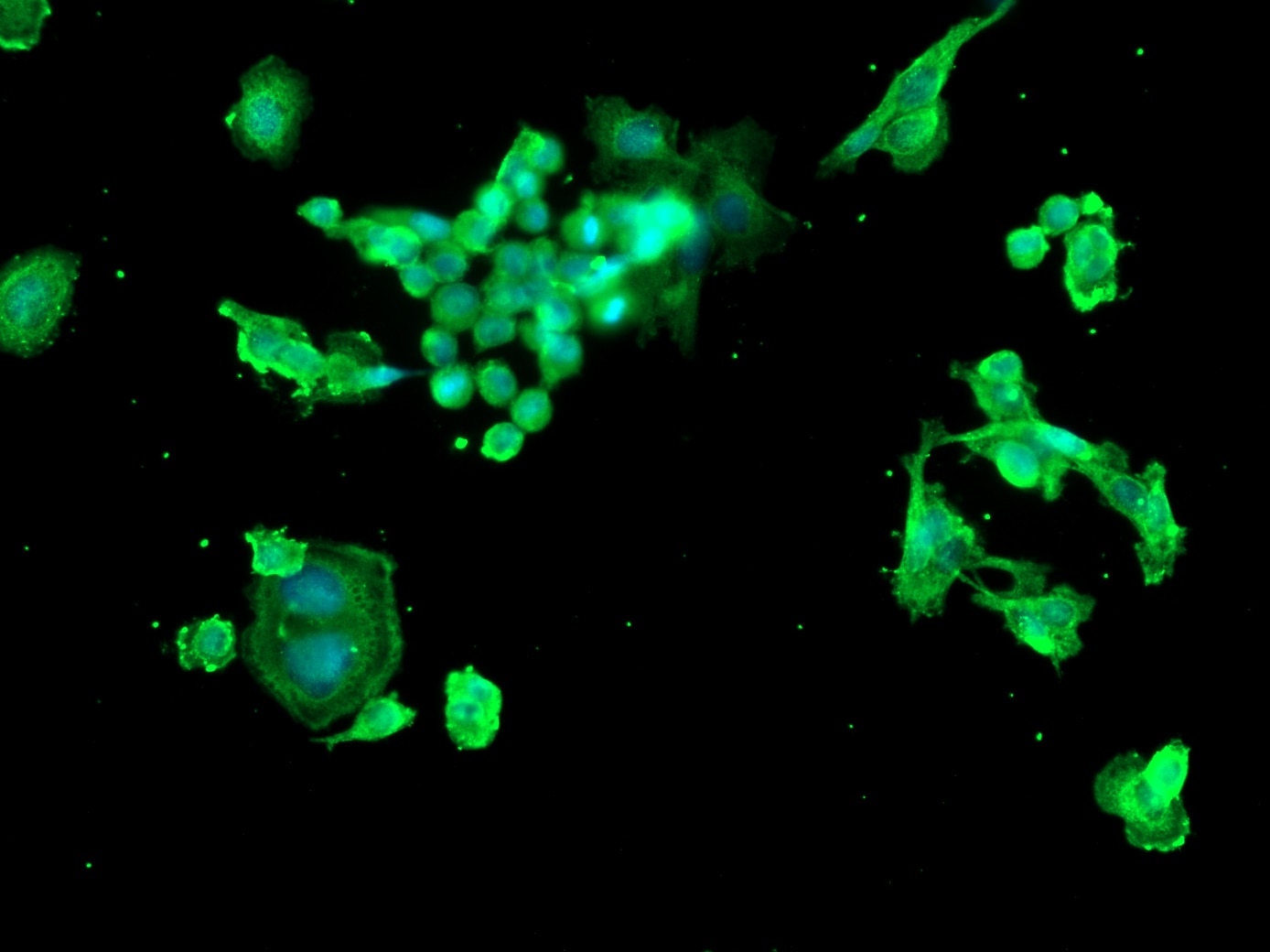
 fixed HeLa cells using 66248-1-Ig(PD-L1/CD274 antibody) at dilution of 1:300 and Alexa Fluor 488-conjugated AffiniPure Goat Anti-Mouse IgG(H+L).")
Applications testées
| Résultats positifs en WB | cellules A375, cellules A549, cellules HepG2, cellules HSC-T6, cellules K-562, cellules RAW 264.7, cellules THP-1, tissu de muscle squelettique humain, tissu placentaire humain, tissu pulmonaire de porc |
| Résultats positifs en IHC | tissu d'amygdalite humain, tissu cardiaque de souris, tissu cardiaque humain, tissu de cancer du poumon humain, tissu placentaire humain il est suggéré de démasquer l'antigène avec un tampon de TE buffer pH 9.0; (*) À défaut, 'le démasquage de l'antigène peut être 'effectué avec un tampon citrate pH 6,0. |
| Résultats positifs en IF-P | tissu placentaire humain, |
| Résultats positifs en IF/ICC | cellules HeLa, |
Dilution recommandée
| Application | Dilution |
|---|---|
| Western Blot (WB) | WB : 1:2000-1:10000 |
| Immunohistochimie (IHC) | IHC : 1:5000-1:20000 |
| Immunofluorescence (IF)-P | IF-P : 1:400-1:1600 |
| Immunofluorescence (IF)/ICC | IF/ICC : 1:50-1:500 |
| It is recommended that this reagent should be titrated in each testing system to obtain optimal results. | |
| Sample-dependent, check data in validation data gallery | |
Informations sur le produit
66248-1-Ig cible PD-L1/CD274 dans les applications de WB, IHC, IF/ICC, IF-P, IP, CoIP, ChIP, ELISA et montre une réactivité avec des échantillons Humain, porc, rat, souris
| Réactivité | Humain, porc, rat, souris |
| Réactivité citée | rat, Humain, porc, souris |
| Hôte / Isotype | Mouse / IgG1 |
| Clonalité | Monoclonal |
| Type | Anticorps |
| Immunogène | PD-L1/CD274 Protéine recombinante Ag12443 |
| Nom complet | CD274 molecule |
| Masse moléculaire calculée | 290 aa, 33 kDa |
| Poids moléculaire observé | 45-50 kDa, 33 kDa |
| Numéro d’acquisition GenBank | BC074984 |
| Symbole du gène | PD-L1 |
| Identification du gène (NCBI) | 29126 |
| Conjugaison | Non conjugué |
| Forme | Liquide |
| Méthode de purification | Purification par protéine G |
| Tampon de stockage | PBS with 0.02% sodium azide and 50% glycerol |
| Conditions de stockage | Stocker à -20°C. Stable pendant un an après l'expédition. L'aliquotage n'est pas nécessaire pour le stockage à -20oC Les 20ul contiennent 0,1% de BSA. |
Informations générales
PD-L1, also known as CD274 or B7H1, stands for programmed cell death ligand 1. It is a type I transmembrane protein that is thought to repress immune responses by binding to its receptor (PD1), thus inhibiting T-cell activation, proliferation, and cytokine production. It contains V-like and C-like immunoglobulin domains. PD-L1 expression is regulated by various cytokines, such as TNF-α or LPS (ISSN: 1848-7718). Increased expression of this protein in certain types of cancers, e.g., renal cell carcinoma or colon cancer, correlates with poor prognosis.
What is the molecular weight of PD-L1?
Depending on the isoform, the calculated molecular weight of the protein varies between 20 and 33 kDa (176-290 aa).
What are the isoforms of PD-L1?
According to NCBI, three different isoforms have been identified. There are significant differences in the untranslated and protein coding regions.
What is the subcellular localization and tissue specificity of PD-L1?
It is predicted to localize in the plasma membrane of various cell types, with a particularly high expression in placental trophoblast and subsets of immune cells. High levels of PD-L1 protein have also been detected in lung and colon tissues.
What is the function of PD-L1 in immune responses?
PD-L1 is critical for the induction and maintenance of immune self-tolerance during infection or inflammation in normal tissues. The interaction of PD-L1 and its receptors is responsible for preventing auto-immune phenotypes and balancing the overall immune response in situations such as pregnancy or tissue allografts. The interaction between PD-L1 and PD-1 or B7.1 starts an inhibitory signaling cascade, which results in the decreased proliferation of antigen-specific T-cells and increased survival of regulatory T-cells (PMID: 15240681).
How can PD-L1's implication in cancer be used as a drug target?
In certain tumors, high expression of PD-L1 serves as a stop-sign to inhibit the recognition of cancer cells by T-cells (PMID: 23087408). The interaction between PD-L1 and its receptors (PD1 and B7.1) is a mechanism for the tumor to evade the host immune response (PMID: 29357948). Several mAbs have been developed to target that interaction and thus prevent the inactivation of cytotoxic T-cells by the tumor (PMIDs: 23890059, 18173375).
Protocole
| Product Specific Protocols | |
|---|---|
| WB protocol for PD-L1/CD274 antibody 66248-1-Ig | Download protocol |
| IHC protocol for PD-L1/CD274 antibody 66248-1-Ig | Download protocol |
| IF protocol for PD-L1/CD274 antibody 66248-1-Ig | Download protocol |
| Standard Protocols | |
|---|---|
| Click here to view our Standard Protocols |
Publications
| Species | Application | Title |
|---|---|---|
Cell Res Targeting ATAD3A-PINK1-mitophagy axis overcomes chemoimmunotherapy resistance by redirecting PD-L1 to mitochondria | ||
Adv Mater A Bifunctional Lysosome-Targeting Chimera Nanoplatform for Tumor-Selective Protein Degradation and Enhanced Cancer Immunotherapy | ||
Cell Metab Dual impacts of serine/glycine-free diet in enhancing antitumor immunity and promoting evasion via PD-L1 lactylation | ||
Cancer Cell ADORA1 Inhibition Promotes Tumor Immune Evasion by Regulating the ATF3-PD-L1 Axis. | ||
Cell Metab NAD+ Metabolism Maintains Inducible PD-L1 Expression to Drive Tumor Immune Evasion. |
Avis
The reviews below have been submitted by verified Proteintech customers who received an incentive for providing their feedback.
FH Margarita (Verified Customer) (11-20-2024) | Secondary antibody: AF 488
 |
FH hala (Verified Customer) (01-27-2023) | there is 2 bands one at the specific size and the other one is higher
 |
FH Emma (Verified Customer) (11-29-2021) | Used for IF on FFPE prostate tissue at 1:50 with Tris-EDTA antigen retrieval (pH 9.0).
 |
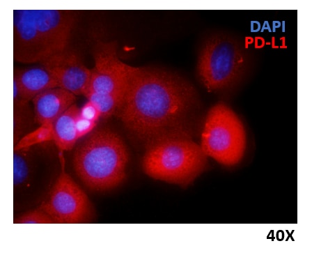
FH Marina (Verified Customer) (06-14-2021) | HCC1937 breast cancer cells were fixed with 4% PFA, permeabilized and stained overnight at 4ºC with anti-PD-L1 Ig diluted 1:300. Nuclei were stained with DAPI.
 |

