- Phare
- Validé par KD/KO
Anticorps Polyclonal de lapin anti-Alix
Alix Polyclonal Antibody for WB, IHC, IF/ICC, IP, ELISA
Hôte / Isotype
Lapin / IgG
Réactivité testée
Humain, rat, souris et plus (4)
Applications
WB, IHC, IF/ICC, IP, ELISA
Conjugaison
Non conjugué
N° de cat : 12422-1-AP
Synonymes
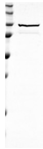
Galerie de données de validation
 at dilution of 1:15000 incubated at room temperature for 1.5 hours.")
 at dilution of 1:10000 incubated at room temperature for 1.5 hours.")
 with sh-Control and sh-Alix transfected HEK-293 cells.")
 at dilution of 1:3000 incubated at room temperature for 1.5 hours.")
 with Jurkat cells lysate 4000ug.")
 at dilution of 1:200 (under 10x lens). Heat mediated antigen retrieval with Tris-EDTA buffer (pH 9.0).")
 at dilution of 1:3000 (under 20x lens). Heat mediated antigen retrieval with Tris-EDTA buffer (pH 9.0).")
 fixed HeLa cells using Alix antibody (12422-1-AP) at dilution of 1:400 and CoraLite®488-Conjugated AffiniPure Goat Anti-Rabbit IgG(H+L).")
Applications testées
| Résultats positifs en WB | cellules HEK-293, cellules HeLa, cellules Jurkat, cellules NIH/3T3, tissu hépatique de rat, tissu hépatique de souris |
| Résultats positifs en IP | cellules Jurkat |
| Résultats positifs en IHC | tissu de cancer du foie humain, tissu de côlon humain il est suggéré de démasquer l'antigène avec un tampon de TE buffer pH 9.0; (*) À défaut, 'le démasquage de l'antigène peut être 'effectué avec un tampon citrate pH 6,0. |
| Résultats positifs en IF/ICC | cellules HeLa, |
Dilution recommandée
| Application | Dilution |
|---|---|
| Western Blot (WB) | WB : 1:5000-1:50000 |
| Immunoprécipitation (IP) | IP : 0.5-4.0 ug for 1.0-3.0 mg of total protein lysate |
| Immunohistochimie (IHC) | IHC : 1:50-1:500 |
| Immunofluorescence (IF)/ICC | IF/ICC : 1:200-1:800 |
| It is recommended that this reagent should be titrated in each testing system to obtain optimal results. | |
| Sample-dependent, check data in validation data gallery | |
Applications publiées
| KD/KO | See 4 publications below |
| WB | See 250 publications below |
| IHC | See 1 publications below |
| IF | See 13 publications below |
| IP | See 3 publications below |
Informations sur le produit
12422-1-AP cible Alix dans les applications de WB, IHC, IF/ICC, IP, ELISA et montre une réactivité avec des échantillons Humain, rat, souris
| Réactivité | Humain, rat, souris |
| Réactivité citée | rat, Humain, Lapin, porc, singe, souris, Hamster |
| Hôte / Isotype | Lapin / IgG |
| Clonalité | Polyclonal |
| Type | Anticorps |
| Immunogène | Alix Protéine recombinante Ag3074 |
| Nom complet | programmed cell death 6 interacting protein |
| Masse moléculaire calculée | 868 aa, 96 kDa |
| Poids moléculaire observé | 75-100 kDa |
| Numéro d’acquisition GenBank | BC020066 |
| Symbole du gène | Alix |
| Identification du gène (NCBI) | 10015 |
| Conjugaison | Non conjugué |
| Forme | Liquide |
| Méthode de purification | Purification par affinité contre l'antigène |
| Tampon de stockage | PBS with 0.02% sodium azide and 50% glycerol |
| Conditions de stockage | Stocker à -20°C. Stable pendant un an après l'expédition. L'aliquotage n'est pas nécessaire pour le stockage à -20oC Les 20ul contiennent 0,1% de BSA. |
Informations générales
ALG-2-interacting protein 1 (ALIX), also known as AIP1 or Hp95, is encoded by PDCD6IP gene and is involved in cell death through mechanisms involving its binding partner ALG-2 (apoptosis-linked gene-2). ALG-2 is a 22-kDa protein containing five serially repetitive EF-hand structures and is defined as a regulator of calcium-induced apoptosis following endoplasmic reticulum (ER) stress. ALIX interacts with ALG-2 through its C-terminal proline-rich region and participates in formation of multivesicular bodies. Recent finding suggest that ALIX is a critical component of caspase 9 activation and apoptosis triggered by calcium. The alix antibody recognizes an additional band of 75-80 kDa which has also been observed in cells and exosomes.
Protocole
| Product Specific Protocols | |
|---|---|
| WB protocol for Alix antibody 12422-1-AP | Download protocol |
| IHC protocol for Alix antibody 12422-1-AP | Download protocol |
| IF protocol for Alix antibody 12422-1-AP | Download protocol |
| IP protocol for Alix antibody 12422-1-AP | Download protocol |
| Standard Protocols | |
|---|---|
| Click here to view our Standard Protocols |
Publications
| Species | Application | Title |
|---|---|---|
Cell Metab Nicotinamide metabolism face-off between macrophages and fibroblasts manipulates the microenvironment in gastric cancer | ||
Nat Immunol Exosomes mediate the cell-to-cell transmission of IFN-α-induced antiviral activity. | ||
Cell Res Intercellular transfer of activated STING triggered by RAB22A-mediated non-canonical autophagy promotes antitumor immunity | ||
Nat Cell Biol Endosomal membrane tension regulates ESCRT-III-dependent intra-lumenal vesicle formation.
| ||
ACS Nano Mesenchymal Stem Cell-Derived Extracellular Vesicles Attenuate Mitochondrial Damage and Inflammation by Stabilizing Mitochondrial DNA. |
Avis
The reviews below have been submitted by verified Proteintech customers who received an incentive for providing their feedback.
FH Kamal (Verified Customer) (02-15-2024) | Strong bands appeared between 70 and 100 kDa.
|
FH Guorong (Verified Customer) (03-22-2022) | Excellent performance with a specific band of expected size
 |
FH Susmita (Verified Customer) (02-11-2022) | all antibodies from Proteintech works great for me
|
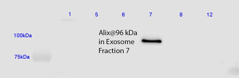
FH Jorge (Verified Customer) (05-15-2019) | Gave a very strong signal using the lowest recommended antibody dilution for Western Blot, diluted it to 1:2000 and worked just as well with a low protein detection substrate. Detected exosomes in fraction 7 of a sucrose gradient as expected.
 |

