- Phare
- Validé par KD/KO
Anticorps Polyclonal de lapin anti-ERp57/ERp60
ERp57/ERp60 Polyclonal Antibody for WB, IHC, IF/ICC, IP, ELISA
Hôte / Isotype
Lapin / IgG
Réactivité testée
Humain, rat, souris et plus (1)
Applications
WB, IHC, IF/ICC, IP, CoIP, ELISA
Conjugaison
Non conjugué
N° de cat : 15967-1-AP
Synonymes
Galerie de données de validation
Applications testées
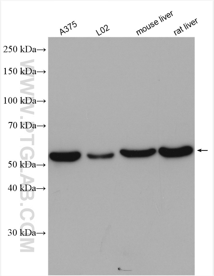
| Résultats positifs en WB | cellules A375, cellules L02, tissu hépatique de rat, tissu hépatique de souris |
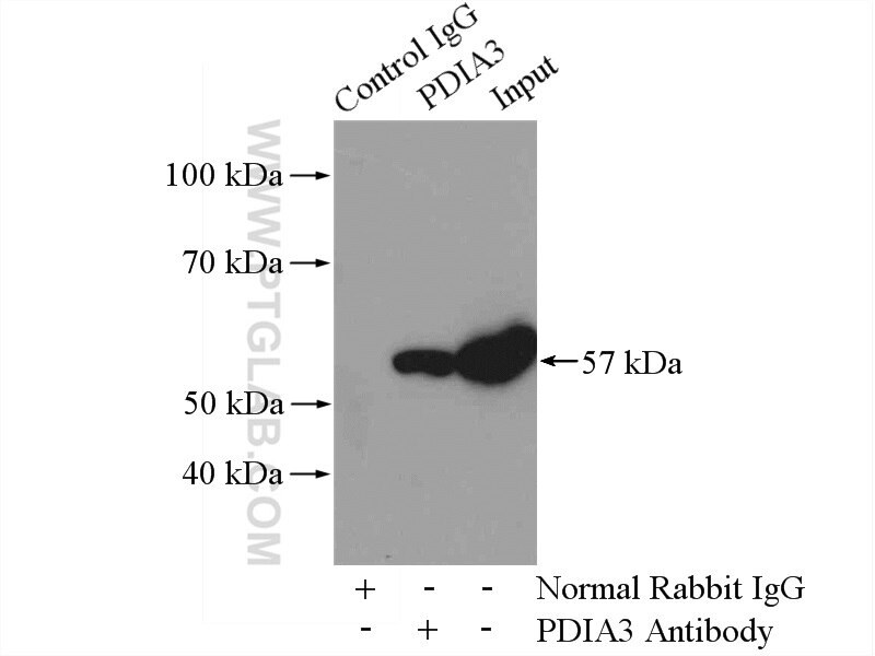
| Résultats positifs en IP | tissu hépatique de souris |
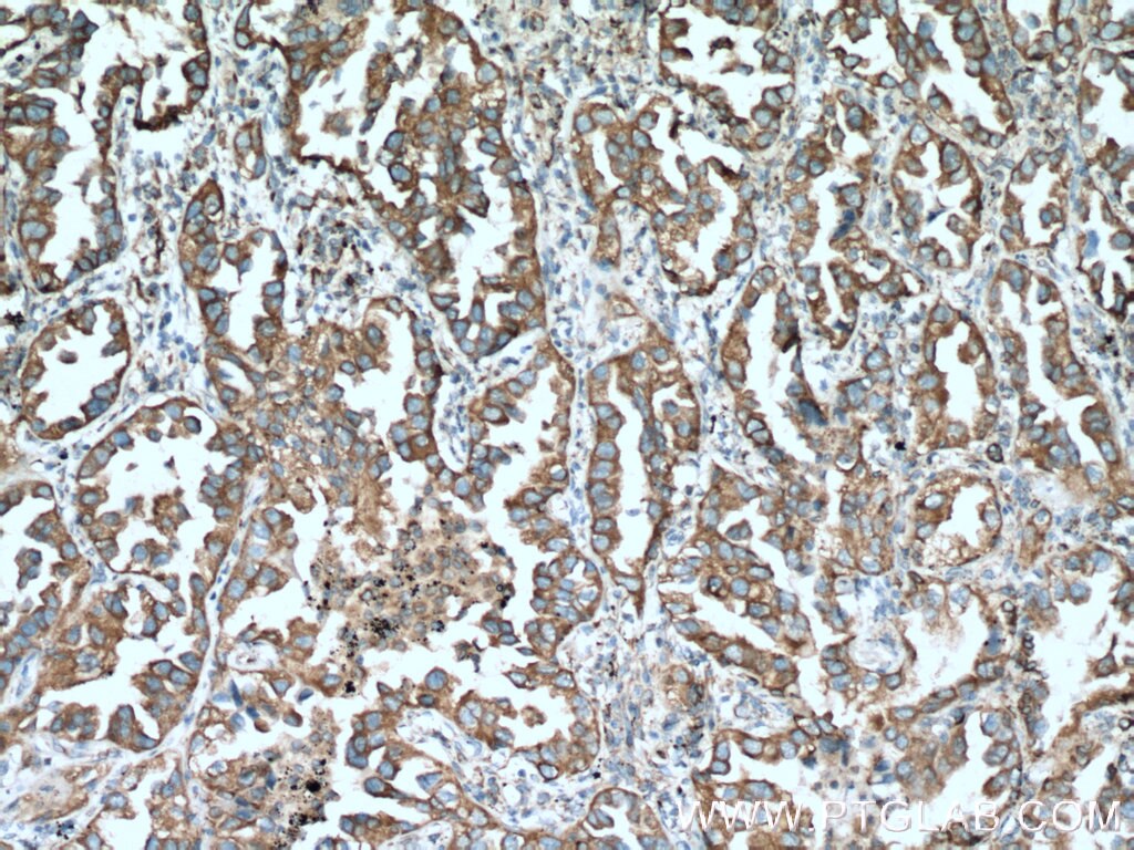
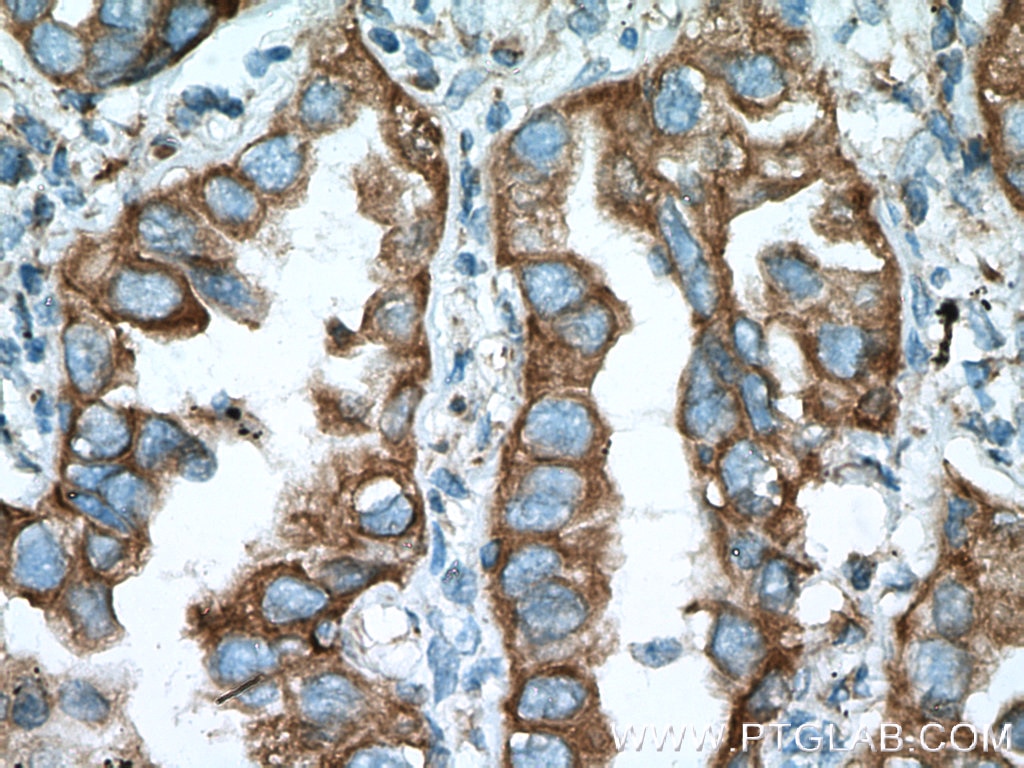
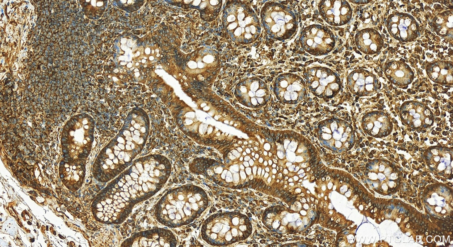
| Résultats positifs en IHC | tissu de cancer du poumon humain, il est suggéré de démasquer l'antigène avec un tampon de TE buffer pH 9.0; (*) À défaut, 'le démasquage de l'antigène peut être 'effectué avec un tampon citrate pH 6,0. |
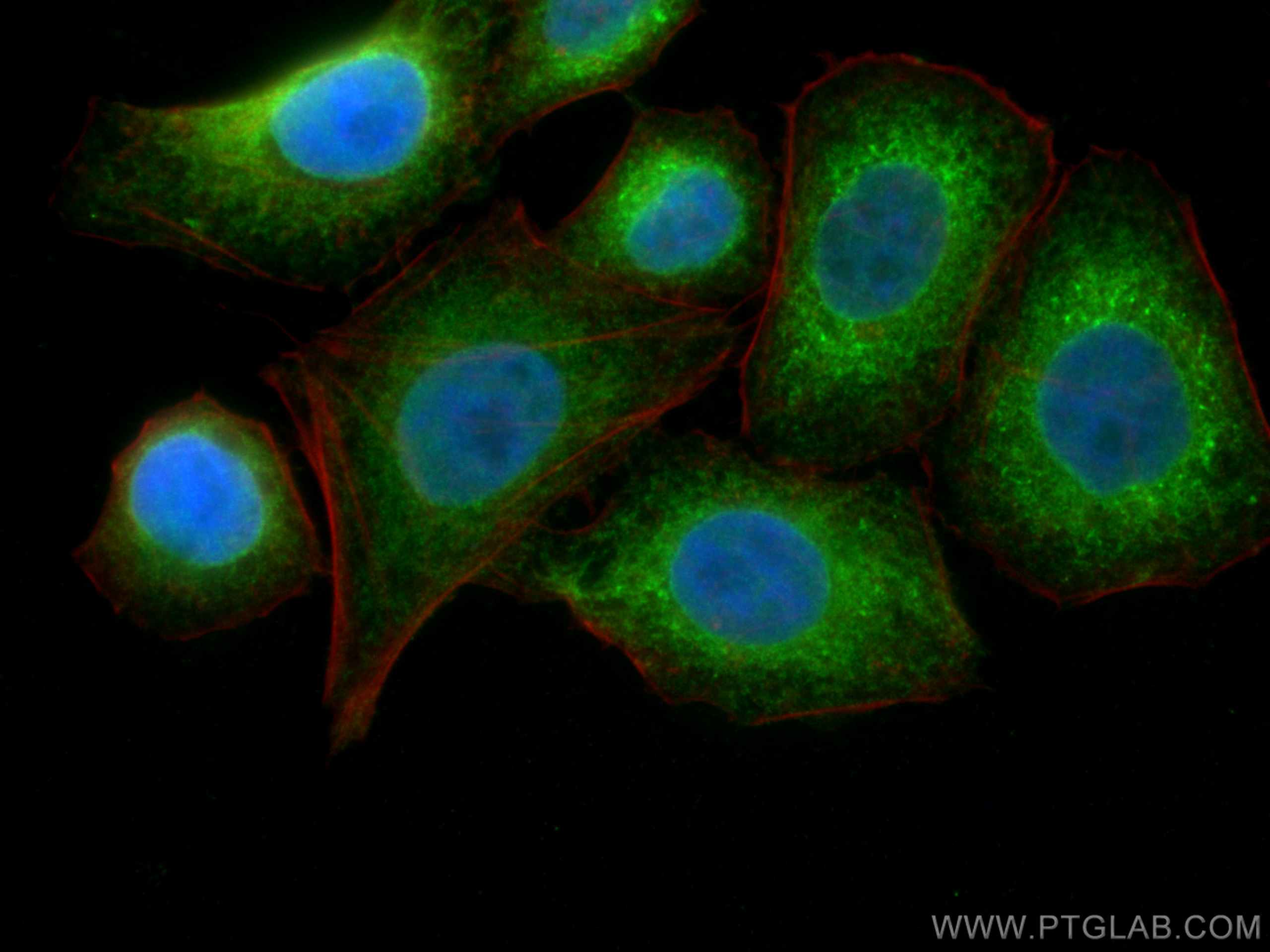
| Résultats positifs en IF/ICC | cellules HepG2 |
Dilution recommandée
| Application | Dilution |
|---|---|
| Western Blot (WB) | WB : 1:2000-1:16000 |
| Immunoprécipitation (IP) | IP : 0.5-4.0 ug for 1.0-3.0 mg of total protein lysate |
| Immunohistochimie (IHC) | IHC : 1:50-1:500 |
| Immunofluorescence (IF)/ICC | IF/ICC : 1:400-1:2000 |
| It is recommended that this reagent should be titrated in each testing system to obtain optimal results. | |
| Sample-dependent, check data in validation data gallery | |
Applications publiées
| KD/KO | See 3 publications below |
| WB | See 33 publications below |
| IHC | See 11 publications below |
| IF | See 14 publications below |
| IP | See 2 publications below |
| ELISA | See 1 publications below |
| CoIP | See 1 publications below |
Informations sur le produit
15967-1-AP cible ERp57/ERp60 dans les applications de WB, IHC, IF/ICC, IP, CoIP, ELISA et montre une réactivité avec des échantillons Humain, rat, souris
| Réactivité | Humain, rat, souris |
| Réactivité citée | rat, Humain, porc, souris |
| Hôte / Isotype | Lapin / IgG |
| Clonalité | Polyclonal |
| Type | Anticorps |
| Immunogène | ERp57/ERp60 Protéine recombinante Ag8741 |
| Nom complet | protein disulfide isomerase family A, member 3 |
| Masse moléculaire calculée | 505 aa, 57 kDa |
| Poids moléculaire observé | 57 kDa |
| Numéro d’acquisition GenBank | BC014433 |
| Symbole du gène | PDIA3 |
| Identification du gène (NCBI) | 2923 |
| Conjugaison | Non conjugué |
| Forme | Liquide |
| Méthode de purification | Purification par affinité contre l'antigène |
| Tampon de stockage | PBS with 0.02% sodium azide and 50% glycerol |
| Conditions de stockage | Stocker à -20°C. Stable pendant un an après l'expédition. L'aliquotage n'est pas nécessaire pour le stockage à -20oC Les 20ul contiennent 0,1% de BSA. |
Informations générales
PDIA3, also named as P58, ER60, ERp57, ERp60, ERp61, GRP57, GRP58 and PI-PLC, is a member of the PDI family, participates in the oxidation, reduction, and isomerization of disulfide bonds for correct folding of secretory proteins before modification and transport in the endoplasmic reticulum. It is associated with apoptosis or inhibition of cancer cell growth. PDIA3 was once thought to be a phospholipase; however, it has been demonstrated that this protein actually has protein disulfide isomerase activity. It is thought that complexes of lectins and PDIA3 mediate protein folding by promoting formation of disulfide bonds in their glycoprotein substrates.
Protocole
| Product Specific Protocols | |
|---|---|
| WB protocol for ERp57/ERp60 antibody 15967-1-AP | Download protocol |
| IHC protocol for ERp57/ERp60 antibody 15967-1-AP | Download protocol |
| IF protocol for ERp57/ERp60 antibody 15967-1-AP | Download protocol |
| IP protocol for ERp57/ERp60 antibody 15967-1-AP | Download protocol |
| Standard Protocols | |
|---|---|
| Click here to view our Standard Protocols |
Publications
| Species | Application | Title |
|---|---|---|
Nat Aging Single-cell and spatial RNA sequencing identify divergent microenvironments and progression signatures in early- versus late-onset prostate cancer | ||
J Control Release A facile and smart strategy to enhance bone regeneration with efficient vitamin D3 delivery through sterosome technology | ||
Mol Biol Evol Long-term artificial selection reveals a role of TCTP in autophagy in mammalian cells. | ||
Redox Biol Protein palmitoylation-mediated palmitic acid sensing causes blood-testis barrier damage via inducing ER stress. | ||
EMBO J TFG clusters COPII-coated transport carriers and promotes early secretory pathway organization. |
Avis
The reviews below have been submitted by verified Proteintech customers who received an incentive for providing their feedback.
FH Prasanna (Verified Customer) (05-07-2018) | Of the four PDIA3/ERp57 antibodies I tried from various companies, this one gave the cleanest and strongest signal by western blot. I was also able to probe multiple blots with the diluted antibody. I will be always come back to this antibody.
|
