- Phare
- Validé par KD/KO
Anticorps Polyclonal de lapin anti-PINK1
PINK1 Polyclonal Antibody for WB, IHC, IF-P, ELISA
Hôte / Isotype
Lapin / IgG
Réactivité testée
Humain, rat, souris et plus (6)
Applications
WB, IHC, IF-P, IP, CoIP, ChIP, ELISA
Conjugaison
Non conjugué
N° de cat : 23274-1-AP
Synonymes
Galerie de données de validation
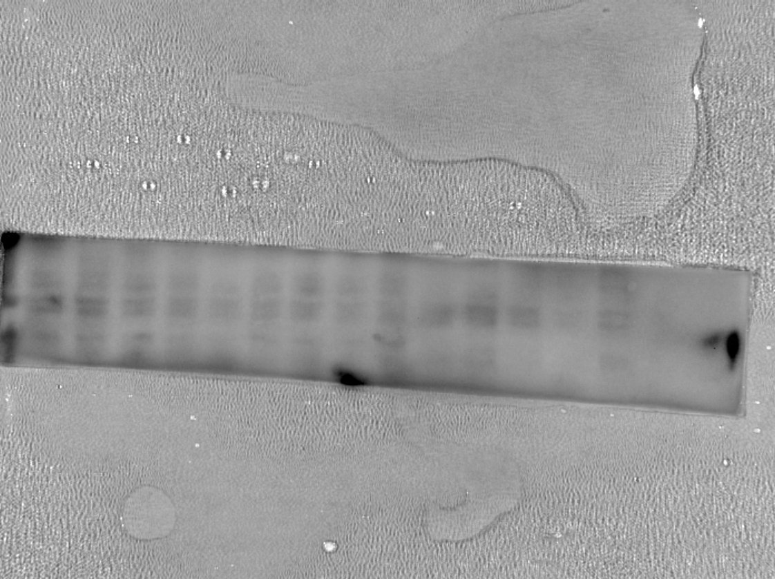
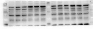
 at dilution of 1:2000 incubated at room temperature for 1.5 hours.")
 at dilution of 1:600 incubated at room temperature for 1.5 hours.")
 at dilution of 1:1000 incubated at room temperature for 1.5 hours.")
 with si-Control and si-PINK1 transfected HeLa cells.")
 at dilution of 1:2000 (under 10x lens). Heat mediated antigen retrieval with Tris-EDTA buffer (pH 9.0).")
 at dilution of 1:2000 (under 40x lens). Heat mediated antigen retrieval with Tris-EDTA buffer (pH 9.0).")
 fixed mouse brain tissue using PINK1 antibody (23274-1-AP) at dilution of 1:400 and CoraLite®488-Conjugated AffiniPure Goat Anti-Rabbit IgG(H+L).")
 fixed mouse brain tissue using PINK1 antibody (23274-1-AP) at dilution of 1:400 and CoraLite®488-Conjugated AffiniPure Goat Anti-Rabbit IgG(H+L).")
 fixed rat brain tissue using PINK1 antibody (23274-1-AP) at dilution of 1:400 and CoraLite®488-Conjugated AffiniPure Goat Anti-Rabbit IgG(H+L).")
 fixed rat brain tissue using PINK1 antibody (23274-1-AP) at dilution of 1:400 and CoraLite®488-Conjugated AffiniPure Goat Anti-Rabbit IgG(H+L).")
Applications testées
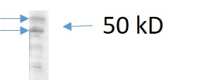
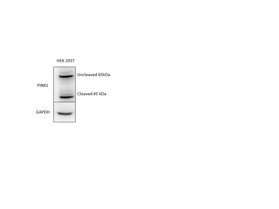
| Résultats positifs en WB | cellules HEK-293, cellules HeLa, cellules PC-12 |
| Résultats positifs en IHC | tissu cérébral de souris, il est suggéré de démasquer l'antigène avec un tampon de TE buffer pH 9.0; (*) À défaut, 'le démasquage de l'antigène peut être 'effectué avec un tampon citrate pH 6,0. |
| Résultats positifs en IF-P | tissu cérébral de souris, tissu cérébral de rat |
Dilution recommandée
| Application | Dilution |
|---|---|
| Western Blot (WB) | WB : 1:1000-1:4000 |
| Immunohistochimie (IHC) | IHC : 1:1000-1:4000 |
| Immunofluorescence (IF)-P | IF-P : 1:200-1:800 |
| It is recommended that this reagent should be titrated in each testing system to obtain optimal results. | |
| Sample-dependent, check data in validation data gallery | |
Informations sur le produit
23274-1-AP cible PINK1 dans les applications de WB, IHC, IF-P, IP, CoIP, ChIP, ELISA et montre une réactivité avec des échantillons Humain, rat, souris
| Réactivité | Humain, rat, souris |
| Réactivité citée | rat, Chèvre, Humain, Lapin, poisson-zèbre, porc, singe, souris, duck |
| Hôte / Isotype | Lapin / IgG |
| Clonalité | Polyclonal |
| Type | Anticorps |
| Immunogène | PINK1 Protéine recombinante Ag19825 |
| Nom complet | PTEN induced putative kinase 1 |
| Masse moléculaire calculée | 581 aa, 63 kDa |
| Poids moléculaire observé | 65 kDa, 45 kDa |
| Numéro d’acquisition GenBank | BC028215 |
| Symbole du gène | PINK1 |
| Identification du gène (NCBI) | 65018 |
| Conjugaison | Non conjugué |
| Forme | Liquide |
| Méthode de purification | Purifié par affinité contre l'antigène |
| Tampon de stockage | PBS with 0.02% sodium azide and 50% glycerol |
| Conditions de stockage | Stocker à -20°C. Stable pendant un an après l'expédition. L'aliquotage n'est pas nécessaire pour le stockage à -20oC Les 20ul contiennent 0,1% de BSA. |
Informations générales
PINK1 is a mitochondrial serine/threonine-protein kinase that protects cells from stress-induced mitochondrial dysfunction. The precursor of PINK1 (65 kDa) is synthesized in the cytosol and is imported into the outer membrane of mitochondria. PINK1 is further transferred into the inner membrane. The full-length PINK1 can be proteolytically processed into 52-55 kDa and 45-46 kDa forms (PMID: 18221368; 25108683; 18031932). The half-life of the mature form of PINK1 is very short and it was proposed that the proteasome is involved in its degradation (PMID: 23472196). The gene of PINK1 maps to chromosome 1p36.12. Two alternatively spliced variants exist, the shorter isoform (30 kDa) produced by alternative splicing. Mutations in the PINK1 gene cause autosomal recessive early-onset Parkinson's disease.
Protocole
| Product Specific Protocols | |
|---|---|
| WB protocol for PINK1 antibody 23274-1-AP | Download protocol |
| IHC protocol for PINK1 antibody 23274-1-AP | Download protocol |
| IF protocol for PINK1 antibody 23274-1-AP | Download protocol |
| Standard Protocols | |
|---|---|
| Click here to view our Standard Protocols |
Publications
| Species | Application | Title |
|---|---|---|
Nat Cell Biol Ammonia-induced lysosomal and mitochondrial damage causes cell death of effector CD8+ T cells | ||
Adv Sci (Weinh) WAC Facilitates Mitophagy-mediated MSC Osteogenesis and New Bone Formation via Protecting PINK1 from Ubiquitination-Dependent Degradation | ||
J Hazard Mater Low-dose deoxynivalenol exposure inhibits hepatic mitophagy and hesperidin reverses this phenomenon by activating SIRT1 | ||
Nat Commun Disuse-associated loss of the protease LONP1 in muscle impairs mitochondrial function and causes reduced skeletal muscle mass and strength. |
Avis
The reviews below have been submitted by verified Proteintech customers who received an incentive for providing their feedback.
FH Javier (Verified Customer) (09-09-2025) | Very good for Western Blotting.
|
FH aidan (Verified Customer) (04-10-2024) | Good results. Would try higher dilution next time (1:500) on precast gel. Used handcast gel overnight incubation at 4 degrees. Blocked 1 hr 5% milk
 |
FH Nin (Verified Customer) (02-27-2023) | It is okay to probe PINK1 with two bands although there are some weak non-specific bands.
 |
FH Yahir Alberto (Verified Customer) (11-12-2021) | The antibody works fine with overnight incubation at 4 °C.
 |
FH Tanusree (Verified Customer) (12-03-2019) | This antibody works good in western blotting analysis using mouse tissues.
|
FH HONGXUE (Verified Customer) (08-19-2019) | The specific is not good. You can detect many bands using WB.
 |

