Anticorps Polyclonal de lapin anti-PLEC
PLEC Polyclonal Antibody for WB, IHC, IF/ICC, ELISA
Hôte / Isotype
Lapin / IgG
Réactivité testée
Humain, souris
Applications
WB, IHC, IF/ICC, CoIP, ELISA
Conjugaison
Non conjugué
N° de cat : 29170-1-AP
Synonymes
Galerie de données de validation
 at dilution of 1:8000 incubated at room temperature for 1.5 hours.")
 at dilution of 1:2000 (under 40x lens). Heat mediated antigen retrieval with Tris-EDTA buffer (pH 9.0).")
 fixed A549 cells using PLEC antibody (29170-1-AP) at dilution of 1:200 and CoraLite®488-Conjugated AffiniPure Goat Anti-Rabbit IgG(H+L) (SA00013-2).")
Applications testées
| Résultats positifs en WB | cellules HEK-293, tissu cérébral de souris, tissu rénal de souris |
| Résultats positifs en IHC | tissu cardiaque de souris, il est suggéré de démasquer l'antigène avec un tampon de TE buffer pH 9.0; (*) À défaut, 'le démasquage de l'antigène peut être 'effectué avec un tampon citrate pH 6,0. |
| Résultats positifs en IF/ICC | cellules A549, |
Dilution recommandée
| Application | Dilution |
|---|---|
| Western Blot (WB) | WB : 1:2000-1:16000 |
| Immunohistochimie (IHC) | IHC : 1:1000-1:4000 |
| Immunofluorescence (IF)/ICC | IF/ICC : 1:50-1:500 |
| It is recommended that this reagent should be titrated in each testing system to obtain optimal results. | |
| Sample-dependent, check data in validation data gallery | |
Applications publiées
| WB | See 1 publications below |
| CoIP | See 1 publications below |
Informations sur le produit
29170-1-AP cible PLEC dans les applications de WB, IHC, IF/ICC, CoIP, ELISA et montre une réactivité avec des échantillons Humain, souris
| Réactivité | Humain, souris |
| Réactivité citée | Humain |
| Hôte / Isotype | Lapin / IgG |
| Clonalité | Polyclonal |
| Type | Anticorps |
| Immunogène | PLEC Protéine recombinante Ag29418 |
| Nom complet | plectin 1, intermediate filament binding protein 500kDa |
| Masse moléculaire calculée | 532 kDa |
| Poids moléculaire observé | 510 kDa |
| Numéro d’acquisition GenBank | NM_201380 |
| Symbole du gène | Plectin |
| Identification du gène (NCBI) | 5339 |
| Conjugaison | Non conjugué |
| Forme | Liquide |
| Méthode de purification | Purification par affinité contre l'antigène |
| Tampon de stockage | PBS with 0.02% sodium azide and 50% glycerol |
| Conditions de stockage | Stocker à -20°C. Stable pendant un an après l'expédition. L'aliquotage n'est pas nécessaire pour le stockage à -20oC Les 20ul contiennent 0,1% de BSA. |
Informations générales
Plectin is a large (≥500-kDa) protein that is normally expressed in various tissues including skin, muscle, and brain. It binds to a number of cytoskeletal proteins including microtubules and intermediate filaments and is involved in establishment and dynamic modulation of the cytoskeletal network. In normal cells, it plays a crucial role in cytoskeleton network organization. In keratinocytes, plectin is concentrated at the basal surface, where it links intermediate filaments to the cytoplasmic domain of transmembrane glycoproteins such as integrin beta-4. (PMID: 24218614, PMID: 28281696, PMID: 23750011)
Protocole
| Product Specific Protocols | |
|---|---|
| WB protocol for PLEC antibody 29170-1-AP | Download protocol |
| IHC protocol for PLEC antibody 29170-1-AP | Download protocol |
| IF protocol for PLEC antibody 29170-1-AP | Download protocol |
| Standard Protocols | |
|---|---|
| Click here to view our Standard Protocols |
Avis
The reviews below have been submitted by verified Proteintech customers who received an incentive for providing their feedback.
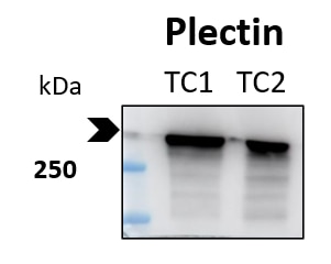
FH Marina (Verified Customer) (03-22-2024) | Samples are two tumour (glioblastoma) cell lines (TC1, TC2). Gradient gel 4-15 %. Dilution 1:2000, primary antibody incubation 4ºC overnight, secondary antibody incubation 1 h room temperature. The blot was incubated with anti-plectin antibody after incubation with anti-spectrin beta chain (ref. 67978-1), stripping and re-blocking. The bands appear at the expected size of the target protein plectin (exposure 2 minutes).
 |

