- Phare
- Validé par KD/KO
Anticorps Polyclonal de lapin anti-PPAR Gamma
PPAR Gamma Polyclonal Antibody for WB, IHC, IF-P, FC (Intra), IP, ELISA
Hôte / Isotype
Lapin / IgG
Réactivité testée
Humain, rat, souris et plus (6)
Applications
WB, IHC, IF-P, FC (Intra), IP, CoIP, CHIP, ELISA
Conjugaison
Non conjugué
N° de cat : 16643-1-AP
Synonymes
Galerie de données de validation
 at dilution of 1:5000 incubated at room temperature for 1.5 hours.")
 at dilution of 1:6000 incubated at room temperature for 1.5 hours.")
 at dilution of 1:2000 incubated at room temperature for 1.5 hours.")
 at dilution of 1:600 incubated at room temperature for 1.5 hours.")
 at dilution of 1:1000 incubated at room temperature for 1.5 hours.")
 with HL-60 cells lysate 2440 ug.")
 at dilution of 1:400 (under 10x lens). Heat mediated antigen retrieval with Tris-EDTA buffer (pH 9.0).")
 at dilution of 1:400 (under 40x lens). Heat mediated antigen retrieval with Tris-EDTA buffer (pH 9.0).")
 at dilution of 1:400 (under 40x lens). Heat mediated antigen retrieval with Tris-EDTA buffer (pH 9.0).")
 at dilution of 1:200 (under 10x lens). Heat mediated antigen retrieval with Tris-EDTA buffer (pH 9.0).")
 at dilution of 1:200 (under 40x lens). Heat mediated antigen retrieval with Tris-EDTA buffer (pH 9.0).")
 at dilution of 1:400 (under 20x lens). Heat mediated antigen retrieval with Tris-EDTA buffer (pH 9.0).")
 at dilution of 1:200 (under 10x lens). Heat mediated antigen retrieval with Tris-EDTA buffer (pH 9.0).")
 at dilution of 1:200 (under 40x lens). Heat mediated antigen retrieval with Tris-EDTA buffer (pH 9.0).")
 at dilution of 1:4000 (under 10x lens). Heat mediated antigen retrieval with Tris-EDTA buffer (pH 9.0).")
 at dilution of 1:4000 (under 40x lens). Heat mediated antigen retrieval with Tris-EDTA buffer (pH 9.0).")
 at dilution of 1:400 (under 10x lens). Heat mediated antigen retrieval with Tris-EDTA buffer (pH 9.0).")
 fixed rat liver tissue using PPAR Gamma antibody (16643-1-AP) at dilution of 1:200 and CoraLite®488-Conjugated AffiniPure Goat Anti-Rabbit IgG(H+L).")
 fixed rat liver tissue using PPAR Gamma antibody (16643-1-AP) at dilution of 1:200 and CoraLite®488-Conjugated AffiniPure Goat Anti-Rabbit IgG(H+L).")
 and CoraLite®488-Conjugated AffiniPure Goat Anti-Rabbit IgG(H+L) at dilution 1:1000 (red), or 0.4 ug Control Antibody. Cells were fixed and permeabilized with Transcription Factor Staining Buffer Kit (PF00011).")
Applications testées
| Résultats positifs en WB | cellules K-562, cellules HL-60, cellules MCF-7, cellules U-937, tissu cardiaque de souris, tissu cardiaque humain |
| Résultats positifs en IP | cellules HL-60, |
| Résultats positifs en IHC | tissu de cancer de la prostate humain, tissu de cancer de la thyroïde humain, tissu de cancer du côlon humain, tissu de cancer du sein humain, tissu placentaire humain il est suggéré de démasquer l'antigène avec un tampon de TE buffer pH 9.0; (*) À défaut, 'le démasquage de l'antigène peut être 'effectué avec un tampon citrate pH 6,0. |
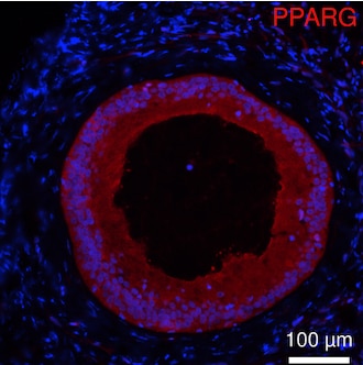
| Résultats positifs en IF-P | tissu hépatique de rat, |
| Résultats positifs en FC (Intra) | cellules HeLa |
Dilution recommandée
| Application | Dilution |
|---|---|
| Western Blot (WB) | WB : 1:1000-1:5000 |
| Immunoprécipitation (IP) | IP : 0.5-4.0 ug for 1.0-3.0 mg of total protein lysate |
| Immunohistochimie (IHC) | IHC : 1:200-1:800 |
| Immunofluorescence (IF)-P | IF-P : 1:50-1:500 |
| Flow Cytometry (FC) (INTRA) | FC (INTRA) : 0.40 ug per 10^6 cells in a 100 µl suspension |
| It is recommended that this reagent should be titrated in each testing system to obtain optimal results. | |
| Sample-dependent, check data in validation data gallery | |
Informations sur le produit
16643-1-AP cible PPAR Gamma dans les applications de WB, IHC, IF-P, FC (Intra), IP, CoIP, CHIP, ELISA et montre une réactivité avec des échantillons Humain, rat, souris
| Réactivité | Humain, rat, souris |
| Réactivité citée | rat, Chèvre, Humain, Lapin, poisson-zèbre, poulet, singe, souris, Hamster |
| Hôte / Isotype | Lapin / IgG |
| Clonalité | Polyclonal |
| Type | Anticorps |
| Immunogène | PPAR Gamma Protéine recombinante Ag10005 |
| Nom complet | peroxisome proliferator-activated receptor gamma |
| Masse moléculaire calculée | 58 kDa |
| Poids moléculaire observé | 50-60 kDa |
| Numéro d’acquisition GenBank | BC006811 |
| Symbole du gène | PPARG |
| Identification du gène (NCBI) | 5468 |
| Conjugaison | Non conjugué |
| Forme | Liquide |
| Méthode de purification | Purification par affinité contre l'antigène |
| Tampon de stockage | PBS with 0.02% sodium azide and 50% glycerol |
| Conditions de stockage | Stocker à -20°C. Stable pendant un an après l'expédition. L'aliquotage n'est pas nécessaire pour le stockage à -20oC Les 20ul contiennent 0,1% de BSA. |
Informations générales
Peroxisome Proliferator-Activated Receptors (PPARs) are ligand-activated intracellular transcription factors, members of the nuclear hormone receptor superfamily (NR), that includes estrogen, thyroid hormone receptors, retinoic acid, Vitamin D3 as well as retinoid X receptors (RXRs). The PPAR subfamily consists of three subtypes encoded by distinct genes denoted PPARα (NR1C1), PPARβ/δ (NR1C2) and PPARγ (NR1C3), which are activated by selective ligands. PPARγ, also named as PPARG, contains one nuclear receptor DNA-binding domain and is a receptor that binds peroxisome proliferators such as hypolipidemic drugs and fatty acids. It plays an important role in the regulation of lipid homeostasis, adipogenesis, ins resistance, and development of various organs. Defects in PPARG are the cause of familial partial lipodystrophy type 3 (FPLD3) and may be associated with susceptibility to obesity. Defects in PPARG can lead to type 2 ins-resistant diabetes and hypertension. PPARG mutations may be associated with colon cancer. Genetic variations in PPARG are associated with susceptibility to glioma type 1 (GLM1). PPARG has two isoforms with molecular weight 57 kDa and 54 kDa (PMID: 9831621), but modified PPARG is about 67 KDa (PMID: 16809887). PPARG2 is a splice variant and has an additional 30 amino acids at the N-terminus (PMID: 15689403). Experimental data indicate that a 45 kDa protein displaying three different sequences immunologically related to the nuclear receptor PPARG2 is located in mitochondria (mt-PPAR). However, the molecular weight of this protein is clearly less when compared to that of PPARG2 (57 kDa) (PMID: 10922459). PPARG has been reported to be localized mainly (but not always) in the nucleus. PPARG can also be detected in the cytoplasm and was reported to possess extra-nuclear/non-genomic actions (PMID: 17611413; 19432669; 14681322).
Protocole
| Product Specific Protocols | |
|---|---|
| WB protocol for PPAR Gamma antibody 16643-1-AP | Download protocol |
| IHC protocol for PPAR Gamma antibody 16643-1-AP | Download protocol |
| IF protocol for PPAR Gamma antibody 16643-1-AP | Download protocol |
| IP protocol for PPAR Gamma antibody 16643-1-AP | Download protocol |
| FC protocol for PPAR Gamma antibody 16643-1-AP | Download protocol |
| Standard Protocols | |
|---|---|
| Click here to view our Standard Protocols |
Publications
| Species | Application | Title |
|---|---|---|
Nat Nanotechnol Photoacoustic molecular imaging-escorted adipose photodynamic-browning synergy for fighting obesity with virus-like complexes. | ||
ACS Nano Dual Inhibition of Endoplasmic Reticulum Stress and Oxidation Stress Manipulates the Polarization of Macrophages under Hypoxia to Sensitize Immunotherapy. | ||
Adv Sci (Weinh) Photothermal-Responsive Soluble Microneedle Patches for Meibomian Gland Dysfunction Therapy | ||
J Pineal Res Single-cell RNA sequencing of preadipocytes reveals the cell fate heterogeneity induced by melatonin. | ||
Nat Commun N1-methyladenosine methylation in tRNA drives liver tumourigenesis by regulating cholesterol metabolism. | ||
Nat Commun METTL3 is essential for postnatal development of brown adipose tissue and energy expenditure in mice. |
Avis
The reviews below have been submitted by verified Proteintech customers who received an incentive for providing their feedback.
FH Tianyi (Verified Customer) (08-30-2024) | Antigen retrieval in Tris/EDTA buffer and incubated with AF594 at room temperature for 2 hours.
 |
FH Surendra (Verified Customer) (12-30-2023) | I purchased this antibody by seeing single band on web page, but it is giving several bands, so its really tough to determine which band is real PPAR gamma.
|
FH Giulia (Verified Customer) (02-21-2022) | The antibody performed well
|
FH Boyan (Verified Customer) (04-24-2019) | By WB, this antibody labeled many bands, which makes it very hard to distinguish which one is the actual PPARg.
|

