- Phare
- Validé par KD/KO
Anticorps Polyclonal de lapin anti-COX2/ Cyclooxygenase 2/ PTGS2
COX2/ Cyclooxygenase 2/ PTGS2 Polyclonal Antibody for WB, IHC, IF/ICC, ELISA
Hôte / Isotype
Lapin / IgG
Réactivité testée
Humain, souris et plus (3)
Applications
WB, IHC, IF/ICC, CoIP, ELISA
Conjugaison
Non conjugué
N° de cat : 12375-1-AP
Synonymes
Galerie de données de validation
 at dilution of 1:2000 incubated at room temperature for 1.5 hours.")
 at dilution of 1:3000 incubated at room temperature for 1.5 hours.")
 at dilution of 1:1500 incubated at room temperature for 1.5 hours.")
 at dilution of 1:500 incubated at room temperature for 1.5 hours.")
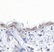
 at dilution of 1:200 (under 10x lens). Heat mediated antigen retrieval with Tris-EDTA buffer (pH 9.0).")
 at dilution of 1:200 (under 40x lens). Heat mediated antigen retrieval with Tris-EDTA buffer (pH 9.0).")
 at dilution of 1:400 (under 20x lens). Heat mediated antigen retrieval with Tris-EDTA buffer (pH 9.0).")
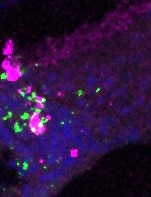
 fixed A549 cells using COX2/ Cyclooxygenase 2/ PTGS2 antibody (12375-1-AP) at dilution of 1:400 and CoraLite®488-Conjugated AffiniPure Goat Anti-Rabbit IgG(H+L) (SA00013-2).")
Applications testées
| Résultats positifs en WB | cellules A549, cellules HEK-293, cellules HeLa, cellules NIH/3T3, cellules Raji, cellules RAW 264.7 |
| Résultats positifs en IHC | tissu de cancer du sein humain, il est suggéré de démasquer l'antigène avec un tampon de TE buffer pH 9.0; (*) À défaut, 'le démasquage de l'antigène peut être 'effectué avec un tampon citrate pH 6,0. |
| Résultats positifs en IF/ICC | cellules A549, |
Dilution recommandée
| Application | Dilution |
|---|---|
| Western Blot (WB) | WB : 1:1000-1:4000 |
| Immunohistochimie (IHC) | IHC : 1:50-1:500 |
| Immunofluorescence (IF)/ICC | IF/ICC : 1:200-1:800 |
| It is recommended that this reagent should be titrated in each testing system to obtain optimal results. | |
| Sample-dependent, check data in validation data gallery | |
Applications publiées
| KD/KO | See 1 publications below |
| WB | See 416 publications below |
| IHC | See 44 publications below |
| IF | See 26 publications below |
| CoIP | See 1 publications below |
Informations sur le produit
12375-1-AP cible COX2/ Cyclooxygenase 2/ PTGS2 dans les applications de WB, IHC, IF/ICC, CoIP, ELISA et montre une réactivité avec des échantillons Humain, souris
| Réactivité | Humain, souris |
| Réactivité citée | rat, Humain, porc, poulet, souris |
| Hôte / Isotype | Lapin / IgG |
| Clonalité | Polyclonal |
| Type | Anticorps |
| Immunogène | COX2/ Cyclooxygenase 2/ PTGS2 Protéine recombinante Ag3025 |
| Nom complet | prostaglandin-endoperoxide synthase 2 (prostaglandin G/H synthase and cyclooxygenase) |
| Masse moléculaire calculée | 604 aa, 68 kDa |
| Poids moléculaire observé | 70-74 kDa |
| Numéro d’acquisition GenBank | BC013734 |
| Symbole du gène | COX2/PTGS2 |
| Identification du gène (NCBI) | 5743 |
| Conjugaison | Non conjugué |
| Forme | Liquide |
| Méthode de purification | Purification par affinité contre l'antigène |
| Tampon de stockage | PBS with 0.02% sodium azide and 50% glycerol |
| Conditions de stockage | Stocker à -20°C. Stable pendant un an après l'expédition. L'aliquotage n'est pas nécessaire pour le stockage à -20oC Les 20ul contiennent 0,1% de BSA. |
Informations générales
COX2 is an enzyme involved in the synthesis of prostaglandins from arachidonic acid.
1. What is the molecular weight of COX2?
The fully N-glycosylated PTGS2 is 72-74 kDa and the aglycosylated is 66 kDa (PMID:19656660). It also
expresses a band of 39 kDa after unspecific cleavage (PMID:17509125). The 50 kDa band of fragmented
PTGS2 has also previously been detected in AD brains (PMID:14724276).
2. Is COX2 post-translationally modified?
COX2 is a subject of post-translational modifications, including phosphorylation, glycosylation, and
s-nitrosylation (PMID: 28939645).
3. What is the difference between COX1, COX2, and COX3?
There are three isoenzymes that have cyclooxygenase activity: COX1, COX2, and COX3. While COX1
is a ubiquitously expressed constitutive enzyme, expression of COX2 is generally low but can be rapidly
induced by various stimuli (as part of infection and inflammatory response) and is controlled by the
transcription factor NFκB. COX3 is an alternative splice variant of COX1 enzyme and is considered
non-functional in humans.
4. I cannot detect COX2 in my sample during western blotting.
Unlike COX1, COX2 expression is inducible by a variety of stimuli including certain growth factors, cytokines,
and proinflammatory stimuli. COX2 basal expression levels may be very low in unstimulated cells. Additionally,
the COX2 half-life time is short and after stimulation, COX2 protein can be quickly degraded to basal levels
(PMID: 10966456), which should be taken into account during experimental design.
5. What is the role of COX2 in cancer?
COX2 is often upregulated in various cancer types and increased expression of COX2 is associated with
greater angiogenesis of solid tumors, increased invasion, and metastasis, as well as decreased host immunity.
6. What is subcellular localization of COX2?
Both COX1 and COX2 are present in the endoplasmic reticulum (ER) and nuclear envelope (PMID: 9545330),
but COX2 has been reported to be more enriched in the nuclear envelope compared to COX1 (PMID: 7738031).
Protocole
| Product Specific Protocols | |
|---|---|
| WB protocol for COX2/ Cyclooxygenase 2/ PTGS2 antibody 12375-1-AP | Download protocol |
| IHC protocol for COX2/ Cyclooxygenase 2/ PTGS2 antibody 12375-1-AP | Download protocol |
| IF protocol for COX2/ Cyclooxygenase 2/ PTGS2 antibody 12375-1-AP | Download protocol |
| Standard Protocols | |
|---|---|
| Click here to view our Standard Protocols |
Publications
| Species | Application | Title |
|---|---|---|
Adv Sci (Weinh) 3D Printing of a Vascularized Mini-Liver Based on the Size-Dependent Functional Enhancements of Cell Spheroids for Rescue of Liver Failure | ||
Sci Adv An annular corneal microneedle patch for minimally invasive ophthalmic drug delivery | ||
Bone Res Piezo1 expression in chondrocytes controls endochondral ossification and osteoarthritis development | ||
Nat Commun BNC1 deficiency-triggered ferroptosis through the NF2-YAP pathway induces primary ovarian insufficiency | ||
EBioMedicine Clearance of apoptotic cells by mesenchymal stem cells contributes to immunosuppression via PGE2. | ||
EBioMedicine Up-regulated FHL2 inhibits ovulation through interacting with androgen receptor and ERK1/2 in polycystic ovary syndrome. |
Avis
The reviews below have been submitted by verified Proteintech customers who received an incentive for providing their feedback.
FH Uthra (Verified Customer) (10-04-2022) | Paraffin-IHC from embedded mouse skin
 |
FH Reema (Verified Customer) (09-07-2022) | The antibody works perfectly with WB
|
FH Lena (Verified Customer) (02-21-2022) | Used for Paraffin-IHC from embedded mouse bones
|
FH Ryan (Verified Customer) (02-28-2019) | Tissue was fixed in PFA and no further antigen retrieval was necessary. Co-localisation in microglia shown with a known marker (magenta).
 |

