- Phare
- Validé par KD/KO
Anticorps Monoclonal anti-PARK2/Parkin
PARK2/Parkin Monoclonal Antibody for WB, IHC, IF/ICC, ELISA
Hôte / Isotype
Mouse / IgG2b
Réactivité testée
Humain, souris et plus (2)
Applications
WB, IHC, IF/ICC, ELISA
Conjugaison
Non conjugué
CloneNo.
2H5A7
N° de cat : 66674-1-Ig
Synonymes
Galerie de données de validation
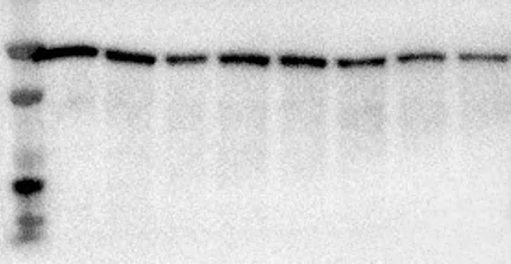
 at dilution of 1:4000 incubated at room temperature for 1.5 hours.")
 at dilution of 1:4000 incubated at room temperature for 1.5 hours.")
 at dilution of 1:4000 incubated at room temperature for 1.5 hours.")
 at dilution of 1:4000 incubated at room temperature for 1.5 hours.")
 at dilution of 1:500 (under 10x lens. Heat mediated antigen retrieval with Tris-EDTA buffer (pH 9.0).")
 at dilution of 1:500 (under 40x lens. Heat mediated antigen retrieval with Tris-EDTA buffer (pH 9.0).")
 fixed RAW 264.7 cells using PARK2/Parkin antibody (66674-1-Ig, Clone: 2H5A7 ) at dilution of 1:400 and CoraLite®488-Conjugated AffiniPure Goat Anti-Mouse IgG(H+L) (SA00013-1), CL594-Phalloidin (red).")
Applications testées
| Résultats positifs en WB | cellules HEK-293, cellules SH-SY5Y, cellules U-251, tissu cérébral de souris |
| Résultats positifs en IHC | tissu cérébral de souris, il est suggéré de démasquer l'antigène avec un tampon de TE buffer pH 9.0; (*) À défaut, 'le démasquage de l'antigène peut être 'effectué avec un tampon citrate pH 6,0. |
| Résultats positifs en IF/ICC | cellules RAW 264.7, |
Dilution recommandée
| Application | Dilution |
|---|---|
| Western Blot (WB) | WB : 1:2000-1:8000 |
| Immunohistochimie (IHC) | IHC : 1:250-1:1000 |
| Immunofluorescence (IF)/ICC | IF/ICC : 1:400-1:1600 |
| It is recommended that this reagent should be titrated in each testing system to obtain optimal results. | |
| Sample-dependent, check data in validation data gallery | |
Applications publiées
| KD/KO | See 1 publications below |
| WB | See 53 publications below |
| IHC | See 6 publications below |
| IF | See 30 publications below |
Informations sur le produit
66674-1-Ig cible PARK2/Parkin dans les applications de WB, IHC, IF/ICC, ELISA et montre une réactivité avec des échantillons Humain, souris
| Réactivité | Humain, souris |
| Réactivité citée | rat, Humain, porc, souris |
| Hôte / Isotype | Mouse / IgG2b |
| Clonalité | Monoclonal |
| Type | Anticorps |
| Immunogène | PARK2/Parkin Protéine recombinante Ag5179 |
| Nom complet | Parkinson disease (autosomal recessive, juvenile) 2, parkin |
| Masse moléculaire calculée | 52 kDa |
| Poids moléculaire observé | 42-52 kDa |
| Numéro d’acquisition GenBank | BC022014 |
| Symbole du gène | Parkin |
| Identification du gène (NCBI) | 5071 |
| Conjugaison | Non conjugué |
| Forme | Liquide |
| Méthode de purification | Purification par protéine A |
| Tampon de stockage | PBS with 0.02% sodium azide and 50% glycerol |
| Conditions de stockage | Stocker à -20°C. Stable pendant un an après l'expédition. L'aliquotage n'est pas nécessaire pour le stockage à -20oC Les 20ul contiennent 0,1% de BSA. |
Informations générales
Parkin, a RING-type E3 ubiquitin-protein ligase, is involved in the ubiquitination pathway and contributes to protection from neurotoxicity induced by unfolded protein stresses. Its ubiquitin-protein ligase activity promotes the degradation of a viariety of proteins including itself. Mutations in Parkin are implicated in the pathogenesis of autosomal recessive familial Parkinson's disease. It has 8 isoforms produced by alternative splicing.
Protocole
| Product Specific Protocols | |
|---|---|
| WB protocol for PARK2/Parkin antibody 66674-1-Ig | Download protocol |
| IHC protocol for PARK2/Parkin antibody 66674-1-Ig | Download protocol |
| IF protocol for PARK2/Parkin antibody 66674-1-Ig | Download protocol |
| Standard Protocols | |
|---|---|
| Click here to view our Standard Protocols |
Publications
| Species | Application | Title |
|---|---|---|
J Pineal Res Melatonin and verteporfin synergistically suppress the growth and stemness of head and neck squamous cell carcinoma through the regulation of mitochondrial dynamics | ||
Autophagy CircEPS15, as a sponge of MIR24-3p ameliorates neuronal damage in Parkinson disease through boosting PINK1-PRKN-mediated mitophagy | ||
Nat Commun Hepatic expression of GAA results in enhanced enzyme bioavailability in mice and non-human primates. | ||
Oxid Med Cell Longev Ginseng-Sanqi-Chuanxiong (GSC) Extracts Ameliorate Diabetes-Induced Endothelial Cell Senescence through Regulating Mitophagy via the AMPK Pathway. | ||
Oxid Med Cell Longev Xuesaitong Combined with Dexmedetomidine Improves Cerebral Ischemia-Reperfusion Injury in Rats by Activating Keap1/Nrf2 Signaling and Mitophagy in Hippocampal Tissue | ||
Cell Death Discov Stomatin-like protein 2 deficiency exacerbates adverse cardiac remodeling |
Avis
The reviews below have been submitted by verified Proteintech customers who received an incentive for providing their feedback.
FH Paula (Verified Customer) (07-26-2024) | Worked well for Western Blotting when made fresh. However, after freezing antibody stock (in 3% BSA) did not work.
 |
FH Tanusree (Verified Customer) (12-03-2019) | This antibody works good in western blotting analysis using mouse tissues.
|

