Anticorps Recombinant de lapin anti-Phospho-TBK1 (Ser172)
Phospho-TBK1 (Ser172) Recombinant Antibody for WB, ELISA
Hôte / Isotype
Lapin / IgG
Réactivité testée
Humain et plus (2)
Applications
WB, ELISA
Conjugaison
Non conjugué
CloneNo.
2K2
N° de cat : 82383-1-RR
Synonymes
Galerie de données de validation
 antibody) at dilution of 1:10000 incubated at room temperature for 1.5 hours. The membrane was stripped and re-blotted with Alpha Tubulin antibody as loading control.")
Applications testées
| Résultats positifs en WB | cellules HeLa traitées à la calyculine A, |
Dilution recommandée
| Application | Dilution |
|---|---|
| Western Blot (WB) | WB : 1:5000-1:50000 |
| It is recommended that this reagent should be titrated in each testing system to obtain optimal results. | |
| Sample-dependent, check data in validation data gallery | |
Applications publiées
| WB | See 7 publications below |
Informations sur le produit
82383-1-RR cible Phospho-TBK1 (Ser172) dans les applications de WB, ELISA et montre une réactivité avec des échantillons Humain
| Réactivité | Humain |
| Réactivité citée | Humain, porc, souris |
| Hôte / Isotype | Lapin / IgG |
| Clonalité | Recombinant |
| Type | Anticorps |
| Immunogène | Peptide |
| Nom complet | TANK-binding kinase 1 |
| Poids moléculaire observé | 84 kDa |
| Numéro d’acquisition GenBank | BC034950 |
| Symbole du gène | TBK1 |
| Identification du gène (NCBI) | 29110 |
| Conjugaison | Non conjugué |
| Forme | Liquide |
| Méthode de purification | Purification par protéine A |
| Tampon de stockage | PBS with 0.02% sodium azide and 50% glycerol |
| Conditions de stockage | Stocker à -20°C. Stable pendant un an après l'expédition. L'aliquotage n'est pas nécessaire pour le stockage à -20oC Les 20ul contiennent 0,1% de BSA. |
Informations générales
TBK1, also named as tumor necrosis factor (TNF) receptor-associated factor NF-kB activator (TANK)-binding kinase 1 (TBK1), NF-kB-activating kinase (NAK), T2K, is a multimeric kinase that modulates inflammation and autophagy. It is a ubiquitously expressed serine-threonine kinase belonging to the 'noncanonical IkB kinases' (IKKs) recognized for its critical role in regulating type I IFN production (PMID: 27211305). And TBK1 is an important player in yet another critical cellular function, autophagy.
Protocole
| Product Specific Protocols | |
|---|---|
| WB protocol for Phospho-TBK1 (Ser172) antibody 82383-1-RR | Download protocol |
| Standard Protocols | |
|---|---|
| Click here to view our Standard Protocols |
Publications
| Species | Application | Title |
|---|---|---|
Signal Transduct Target Ther The cGAS-STING pathway-dependent sensing of mitochondrial DNA mediates ocular surface inflammation | ||
J Virol Pseudorabies virus infection triggers mitophagy to dampen the interferon response and promote viral replication | ||
Mol Med Melatonin alleviates sepsis-induced acute lung injury by inhibiting necroptosis via reducing circulating mtDNA release | ||
Cell Rep Non-canonical isoforms of the mRNA polyadenylation factor WDR33 regulate STING-mediated immune responses | ||
Int J Biol Macromol Inhibiting UGCG prevents PRV infection by decreasing lysosome-associated autophage | ||
Eur J Pharmacol Nitisinone attenuates cartilage degeneration and subchondral osteoclastogenesis in osteoarthritis and concomitantly inhibits the cGAS/STING/NF-κB pathway |
Avis
The reviews below have been submitted by verified Proteintech customers who received an incentive for providing their feedback.
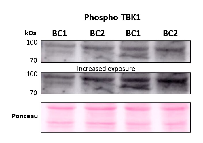
FH Marina (Verified Customer) (09-27-2023) | Western blot using p-TBK1 antibody at 1:5000 dilution overnight at 4ºC. Samples are two breast cancer (BC) cell lines, ran in duplicate. Ponceau staining is shown as total protein loading control. A more prominent band can be seen below 100 kDa, and after more exposure, another band at around 80 kDa can be observed.
 |

