- Phare
- Validé par KD/KO
Anticorps Polyclonal de lapin anti-RAB5A
RAB5A Polyclonal Antibody for WB, IP, IHC, ELISA
Hôte / Isotype
Lapin / IgG
Réactivité testée
Humain, rat, souris et plus (2)
Applications
WB, IHC, IF, IP, ELISA
Conjugaison
Non conjugué
N° de cat : 11947-1-AP
Synonymes
Galerie de données de validation
 at dilution of 1:6000 incubated at room temperature for 1.5 hours.")
 at dilution of 1:10000 incubated at room temperature for 1.5 hours.")
 at dilution of 1:300 incubated at room temperature for 1.5 hours.")
 with mouse brain tissue lysate 1680 ug.")
 at dilution of 1:200 (under 10x lens). Heat mediated antigen retrieval with Tris-EDTA buffer (pH 9.0).")
 at dilution of 1:200 (under 40x lens). Heat mediated antigen retrieval with Tris-EDTA buffer (pH 9.0).")
 at dilution of 1:200 (under 10x lens). Heat mediated antigen retrieval with Tris-EDTA buffer (pH 9.0).")
 at dilution of 1:200 (under 10x lens).")
 at dilution of 1:200 (under 40x lens).")
Applications testées
| Résultats positifs en WB | tissu cérébral de souris, cellules HeLa, tissu cérébral de rat, tissu cérébral humain |
| Résultats positifs en IP | tissu cérébral de souris, |
| Résultats positifs en IHC | tissu de gliome humain, tissu cérébral de souris, tissu pancréatique humain il est suggéré de démasquer l'antigène avec un tampon de TE buffer pH 9.0; (*) À défaut, 'le démasquage de l'antigène peut être 'effectué avec un tampon citrate pH 6,0. |
Dilution recommandée
| Application | Dilution |
|---|---|
| Western Blot (WB) | WB : 1:5000-1:50000 |
| Immunoprécipitation (IP) | IP : 0.5-4.0 ug for 1.0-3.0 mg of total protein lysate |
| Immunohistochimie (IHC) | IHC : 1:50-1:500 |
| It is recommended that this reagent should be titrated in each testing system to obtain optimal results. | |
| Sample-dependent, check data in validation data gallery | |
Applications publiées
| KD/KO | See 8 publications below |
| WB | See 30 publications below |
| IHC | See 7 publications below |
| IF | See 19 publications below |
| IP | See 2 publications below |
Informations sur le produit
11947-1-AP cible RAB5A dans les applications de WB, IHC, IF, IP, ELISA et montre une réactivité avec des échantillons Humain, rat, souris
| Réactivité | Humain, rat, souris |
| Réactivité citée | rat, Humain, porc, singe, souris |
| Hôte / Isotype | Lapin / IgG |
| Clonalité | Polyclonal |
| Type | Anticorps |
| Immunogène | RAB5A Protéine recombinante Ag2549 |
| Nom complet | RAB5A, member RAS oncogene family |
| Masse moléculaire calculée | 215 aa, 24 kDa |
| Poids moléculaire observé | 24 kDa |
| Numéro d’acquisition GenBank | BC001267 |
| Symbole du gène | RAB5A |
| Identification du gène (NCBI) | 5868 |
| Conjugaison | Non conjugué |
| Forme | Liquide |
| Méthode de purification | Purification par affinité contre l'antigène |
| Tampon de stockage | PBS with 0.02% sodium azide and 50% glycerol |
| Conditions de stockage | Stocker à -20°C. Stable pendant un an après l'expédition. L'aliquotage n'est pas nécessaire pour le stockage à -20oC Les 20ul contiennent 0,1% de BSA. |
Protocole
| Product Specific Protocols | |
|---|---|
| WB protocol for RAB5A antibody 11947-1-AP | Download protocol |
| IHC protocol for RAB5A antibody 11947-1-AP | Download protocol |
| IP protocol for RAB5A antibody 11947-1-AP | Download protocol |
| Standard Protocols | |
|---|---|
| Click here to view our Standard Protocols |
Publications
| Species | Application | Title |
|---|---|---|
Autophagy Live imaging of intra-lysosome pH in cell lines and primary neuronal culture using a novel genetically encoded biosensor. | ||
Autophagy RAB7 activity is required for the regulation of mitophagy in oocyte meiosis and oocyte quality control during ovarian aging. | ||
Diabetes Atorvastatin Targets the Islet Mevalonate Pathway to Dysregulate mTOR Signaling and Reduce β-Cell Functional Mass.
| ||
Sci Signal Semaphorin 3A activates the guanosine triphosphatase Rab5 to promote growth cone collapse and organize callosal axon projections. | ||
Elife Capping protein regulates endosomal trafficking by controlling F-actin density around endocytic vesicles and recruiting RAB5 effectors. |
Avis
The reviews below have been submitted by verified Proteintech customers who received an incentive for providing their feedback.
FH Sophy (Verified Customer) (02-27-2023) | The antibody was used at 1:1000 for western blot. The signal showed up but not as strong as expected. But since this was the only antibody purchased for Rab5A, I had no comparison to antibodies to other vendors.
|
FH Xin (Verified Customer) (01-24-2022) | Very good antibody in WB (25 kD) with a high titer
|
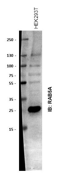
FH Tom (Verified Customer) (11-13-2020) | HEK293T cell extracts (10ug/lane). Primary antibody (1:2000) in block (5% BSA) incubated at 4 degrees overnight. Goat anti-rabbit HRP secondary antibody (1:10,000) incubated for 1 hour at RT.
 |
FH Aamir (Verified Customer) (01-19-2020) | Used in HEK cells. Good signal for WB and IF
|

