- Phare
- Validé par KD/KO
Anticorps Polyclonal de lapin anti-REDD1 specific
REDD1 specific Polyclonal Antibody for WB, IHC, IF/ICC, IP, ELISA
Hôte / Isotype
Lapin / IgG
Réactivité testée
Humain, souris et plus (5)
Applications
WB, IHC, IF/ICC, IP, CoIP, chIP, ELISA
Conjugaison
Non conjugué
N° de cat : 10638-1-AP
Synonymes
Galerie de données de validation
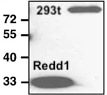
 at dilution of 1:6000 incubated at room temperature for 1.5 hours.")
 with si-control and si-REDD1 transfected PC-3 cells.")
 with sh-Control and sh-REDD1 specific transfected K-562 cells.")
 at dilution of 1:600 incubated at room temperature for 6 hours. .")
 at dilution of 1:1000 incubated at room temperature for 1.5 hours.")
 at dilution of 1:6000 incubated at room temperature for 1.5 hours.")
 at dilution of 1:1000 incubated at room temperature for 1.5 hours.")
 with MCF-7 cells lysate 2500ug.")
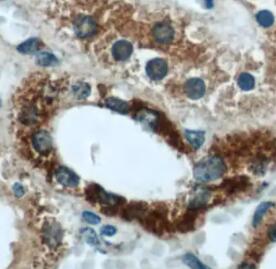
 at dilution of 1:200 (under 40x lens). Heat mediated antigen retrieval with Tris-EDTA buffer (pH 9.0).")
 at dilution of 1:600 (under 40x lens). Heat mediated antigen retrieval with Tris-EDTA buffer (pH 9.0).")
 at dilution of 1:600 (under 40x lens). Heat mediated antigen retrieval with Tris-EDTA buffer (pH 9.0).")
 at dilution of 1:50 (under 10x lens).")
 at dilution of 1:50 (under 40x lens).")
 at dilution of 1:100 (under 10x lens).")
 at dilution of 1:100 (under 40x lens).")
 fixed LNCaP cells using REDD1 specific antibody (10638-1-AP) at dilution of 1:400 and CoraLite®488-Conjugated AffiniPure Goat Anti-Rabbit IgG(H+L).")
Applications testées
| Résultats positifs en WB | cellules A431, cellules A549, cellules DU 145, cellules HeLa traitées au chlorure de cobalt, cellules K-562, cellules LNCaP, cellules PC-3, cellules SH-SY5Y |
| Résultats positifs en IP | cellules MCF-7 |
| Résultats positifs en IHC | tissu de cancer du poumon humain, tissu cardiaque humain, tissu hépatique humain il est suggéré de démasquer l'antigène avec un tampon de TE buffer pH 9.0; (*) À défaut, 'le démasquage de l'antigène peut être 'effectué avec un tampon citrate pH 6,0. |
| Résultats positifs en IF/ICC | cellules LNCaP, |
Dilution recommandée
| Application | Dilution |
|---|---|
| Western Blot (WB) | WB : 1:2000-1:12000 |
| Immunoprécipitation (IP) | IP : 0.5-4.0 ug for 1.0-3.0 mg of total protein lysate |
| Immunohistochimie (IHC) | IHC : 1:50-1:500 |
| Immunofluorescence (IF)/ICC | IF/ICC : 1:200-1:800 |
| It is recommended that this reagent should be titrated in each testing system to obtain optimal results. | |
| Sample-dependent, check data in validation data gallery | |
Informations sur le produit
10638-1-AP cible REDD1 specific dans les applications de WB, IHC, IF/ICC, IP, CoIP, chIP, ELISA et montre une réactivité avec des échantillons Humain, souris
| Réactivité | Humain, souris |
| Réactivité citée | rat, Humain, Lapin, porc, souris, Gerbille, squirrel |
| Hôte / Isotype | Lapin / IgG |
| Clonalité | Polyclonal |
| Type | Anticorps |
| Immunogène | REDD1 specific Protéine recombinante Ag0965 |
| Nom complet | DNA-damage-inducible transcript 4 |
| Masse moléculaire calculée | 25 kDa |
| Poids moléculaire observé | 32-35 kDa |
| Numéro d’acquisition GenBank | BC007714 |
| Symbole du gène | REDD1/DDIT4 |
| Identification du gène (NCBI) | 54541 |
| Conjugaison | Non conjugué |
| Forme | Liquide |
| Méthode de purification | Purification par affinité contre l'antigène |
| Tampon de stockage | PBS with 0.02% sodium azide and 50% glycerol |
| Conditions de stockage | Stocker à -20°C. Stable pendant un an après l'expédition. L'aliquotage n'est pas nécessaire pour le stockage à -20oC Les 20ul contiennent 0,1% de BSA. |
Informations générales
REDD1, also named as RTP801 and DDIT4, belongs to the DDIT4 family. REDD1 promotes neuronal cell death. It is a novel transcriptional target of p53 implicated ROS in the p53-dependent DNA damage response. REDD1 controlled cell growth under energy stress, as an essential regulator of TOR activity through the TSC1/2 complex. REDD-1 expression has also been linked to apoptosis, Aβ toxicity and the pathogenesis of ischemic diseases. As an HIF-1-responsive gene, REDD-1 exhibits strong hypoxia-dependent upregulation in ischemic cells of neuronal origin[PMID: 19996311]. In response to stress due to DNA damage and glucocorticoid treatment, REDD-1 is upregulated at the transcriptional level[PMID: 21733849]. REDD-1 negatively regulates the mammalian target of Rapamycin, a serine/threonine kinase often referred to as mTOR[PMID: 22951983]. It is crucial in the coupling of extra- and intracellular cues to mTOR regulation. The absence of REDD-1 is associated with the development of retinopathy, a major cause of blindness[PMID: 22304497]. REDD1 is a new host defense factor, and chemical activation of REDD1 expression represents a potent antiviral intervention strategy[PMID: 21909097]. The calculated molecular weight of REDD1 is 25 kDa. Because of multiple lysines in the proteins, REDD1 offen migrates around 35 kDa on Western blot[PMID: 19221489]. This antibody is a rabbit polyclonal antibody raised against full length human REDD1 antigen. This antibody is specific to the REDD1 from siRNA experiment (PMID:24713927)
Protocole
| Product Specific Protocols | |
|---|---|
| WB protocol for REDD1 specific antibody 10638-1-AP | Download protocol |
| IHC protocol for REDD1 specific antibody 10638-1-AP | Download protocol |
| IF protocol for REDD1 specific antibody 10638-1-AP | Download protocol |
| IP protocol for REDD1 specific antibody 10638-1-AP | Download protocol |
| Standard Protocols | |
|---|---|
| Click here to view our Standard Protocols |
Publications
| Species | Application | Title |
|---|---|---|
Cell Metab Disrupted methionine cycle triggers muscle atrophy in cancer cachexia through epigenetic regulation of REDD1 | ||
Cancer Cell A UBE2O-AMPKα2 Axis that Promotes Tumor Initiation and Progression Offers Opportunities for Therapy. | ||
Nat Immunol The transcription factor Rfx7 limits metabolism of NK cells and promotes their maintenance and immunity. |
Avis
The reviews below have been submitted by verified Proteintech customers who received an incentive for providing their feedback.
FH Yuki (Verified Customer) (11-10-2022) | This product was used for IHC in human tissue.
 |
FH Stephane (Verified Customer) (11-26-2020) | The antibody performs great on human cell extract. It detects specific bands with the REDD1 band resolving around 30-35 kD.
 |
FH Jamal (Verified Customer) (09-17-2019) | This product was used for Western Blotting in mouse cortex tissue.
|
FH Bradley (Verified Customer) (08-19-2019) | I have used this antibody on numerous occasions with success. The antibody performs great on human cell extract, as evidenced by a single immunoreactive band. The antibody performs moderately well on mouse skeletal muscle extract as it detects many non-specific bands with the REDD1 band resolving around 32-35 kD (specificity determined using REDD1-/- samples). The antibody is capable of detecting large changes in REDD1 protein in mouse skeletal muscle (i.e. induction following dexamethasone treatment), but smaller changes are difficult to assess in this tissue. This antibody will detect smaller changes in human cell lines such as HEK 293 or Hela cells.
|

