- Phare
- Validé par KD/KO
Anticorps Polyclonal de lapin anti-RICTOR
RICTOR Polyclonal Antibody for WB, IHC, IF/ICC, ELISA
Hôte / Isotype
Lapin / IgG
Réactivité testée
Humain, souris et plus (1)
Applications
WB, IHC, IF/ICC, IP, ELISA
Conjugaison
Non conjugué
N° de cat : 27248-1-AP
Synonymes
Galerie de données de validation
 at dilution of 1:4000 incubated at room temperature for 1.5 hours.")
 at dilution of 1:2000 incubated at room temperature for 1.5 hours.")
 at dilution of 1:1000 incubated at room temperature for 1.5 hours.")
 at dilution of 1:200 (under 10x lens. Heat mediated antigen retrieval with Tris-EDTA buffer (pH 9.0).")
 at dilution of 1:200 (under 40x lens. Heat mediated antigen retrieval with Tris-EDTA buffer (pH 9.0).")
 at dilution of 1:600 (under 10x lens). Heat mediated antigen retrieval with Tris-EDTA buffer (pH 9.0).")
 at dilution of 1:600 (under 40x lens). Heat mediated antigen retrieval with Tris-EDTA buffer (pH 9.0).")
 fixed NIH/3T3 cells using RICTOR antibody (27248-1-AP) at dilution of 1:200 and Multi-rAb CoraLite ® Plus 488-Goat Anti-Rabbit Recombinant Secondary Antibody (H+L) (RGAR002).")
 fixed HeLa cells using RICTOR antibody (27248-1-AP) at dilution of 1:400 and Multi-rAb CoraLite ® Plus 488-Goat Anti-Rabbit Recombinant Secondary Antibody (H+L) (RGAR002).")
 fixed HeLa cells using 27248-1-AP (RICTOR antibody) at dilution of 1:50 and CoraLite488-Conjugated AffiniPure Goat Anti-Rabbit IgG(H+L).")
Applications testées
| Résultats positifs en WB | cellules HEK-293, cellules HeLa, cellules HepG2, cellules NIH/3T3 |
| Résultats positifs en IHC | tissu de cancer du poumon humain, tissu testiculaire de souris il est suggéré de démasquer l'antigène avec un tampon de TE buffer pH 9.0; (*) À défaut, 'le démasquage de l'antigène peut être 'effectué avec un tampon citrate pH 6,0. |
| Résultats positifs en IF/ICC | cellules NIH/3T3, cellules HeLa |
Dilution recommandée
| Application | Dilution |
|---|---|
| Western Blot (WB) | WB : 1:1000-1:8000 |
| Immunohistochimie (IHC) | IHC : 1:50-1:500 |
| Immunofluorescence (IF)/ICC | IF/ICC : 1:50-1:500 |
| It is recommended that this reagent should be titrated in each testing system to obtain optimal results. | |
| Sample-dependent, check data in validation data gallery | |
Applications publiées
| KD/KO | See 2 publications below |
| WB | See 22 publications below |
| IHC | See 1 publications below |
| IF | See 6 publications below |
| IP | See 1 publications below |
Informations sur le produit
27248-1-AP cible RICTOR dans les applications de WB, IHC, IF/ICC, IP, ELISA et montre une réactivité avec des échantillons Humain, souris
| Réactivité | Humain, souris |
| Réactivité citée | rat, Humain, souris |
| Hôte / Isotype | Lapin / IgG |
| Clonalité | Polyclonal |
| Type | Anticorps |
| Immunogène | RICTOR Protéine recombinante Ag25649 |
| Nom complet | rapamycin-insensitive companion of mTOR |
| Masse moléculaire calculée | 192 kDa |
| Poids moléculaire observé | 192 kDa |
| Numéro d’acquisition GenBank | BC029608 |
| Symbole du gène | RICTOR |
| Identification du gène (NCBI) | 253260 |
| Conjugaison | Non conjugué |
| Forme | Liquide |
| Méthode de purification | Purification par affinité contre l'antigène |
| Tampon de stockage | PBS with 0.02% sodium azide and 50% glycerol |
| Conditions de stockage | Stocker à -20°C. Stable pendant un an après l'expédition. L'aliquotage n'est pas nécessaire pour le stockage à -20oC Les 20ul contiennent 0,1% de BSA. |
Protocole
| Product Specific Protocols | |
|---|---|
| WB protocol for RICTOR antibody 27248-1-AP | Download protocol |
| IHC protocol for RICTOR antibody 27248-1-AP | Download protocol |
| IF protocol for RICTOR antibody 27248-1-AP | Download protocol |
| Standard Protocols | |
|---|---|
| Click here to view our Standard Protocols |
Publications
| Species | Application | Title |
|---|---|---|
Aging (Albany NY) Interleukin 6 promotes BMP9-induced osteoblastic differentiation through Stat3/mTORC1 in mouse embryonic fibroblasts | ||
Cell Rep Mitochondrial dynamics define muscle fiber type by modulating cellular metabolic pathways | ||
J Cell Physiol Identification of circular dorsal ruffles as signal platforms for the AKT pathway in glomerular podocytes | ||
J Ethnopharmacol Diterpenoid tanshinones inhibit gastric cancer angiogenesis through the PI3K/Akt/mTOR signaling pathway | ||
Cell Commun Signal Rapamycin promotes endothelial-mesenchymal transition during stress-induced premature senescence through the activation of autophagy. |
Avis
The reviews below have been submitted by verified Proteintech customers who received an incentive for providing their feedback.
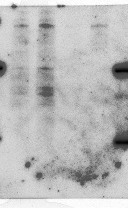
FH Joleen (Verified Customer) (06-12-2019) | The antibody recognizes RICTOR which is around ~200kD. It is the top most band. The first two lanes were cell lysates and show that the antibody recognizes other proteins non-specifically. The last lane contained eluted proteins that are presumably proximal to the cell membrane. RICTOR shows up there cleanly.
 |

