Anticorps Polyclonal de lapin anti-RIPK3
RIPK3 Polyclonal Antibody for WB, IHC, IF/ICC, IP, Indirect ELISA
Hôte / Isotype
Lapin / IgG
Réactivité testée
Humain
Applications
WB, IHC, IF/ICC, IP, Indirect ELISA
Conjugaison
Non conjugué
N° de cat : 17563-1-PBS
Synonymes
Galerie de données de validation
Informations sur le produit
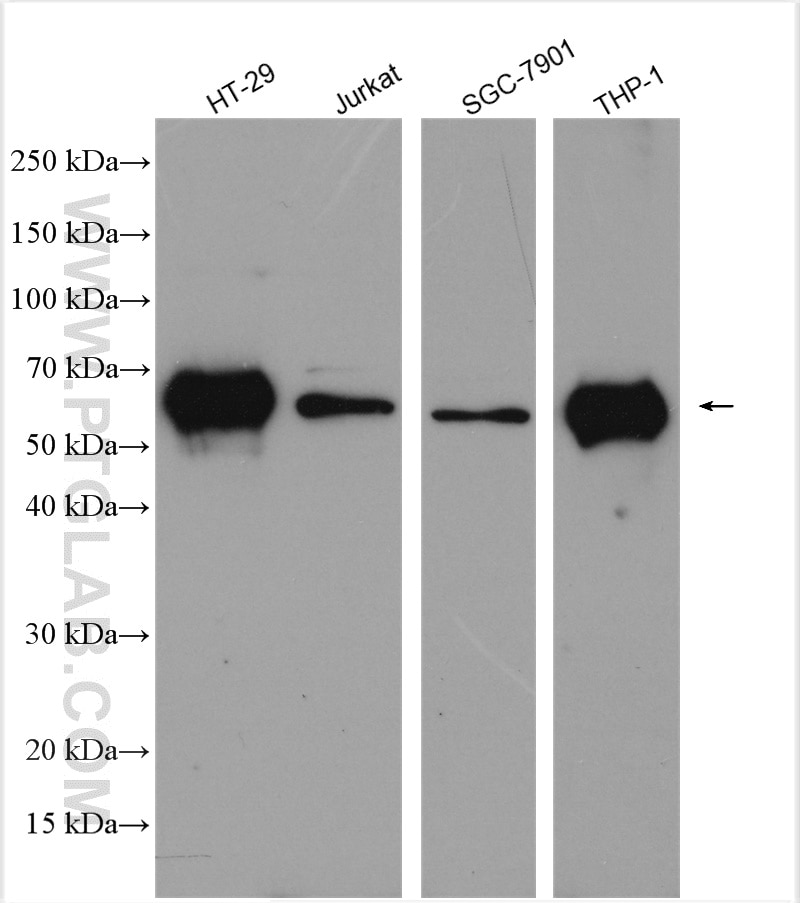
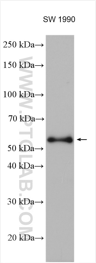
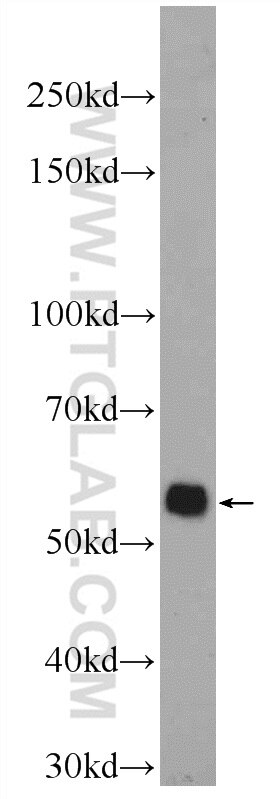
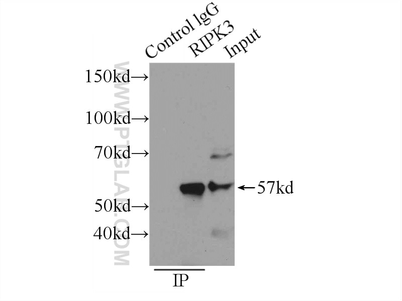
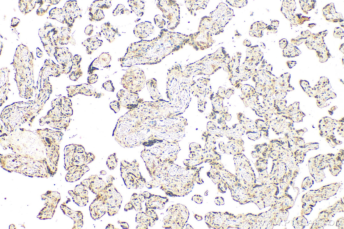
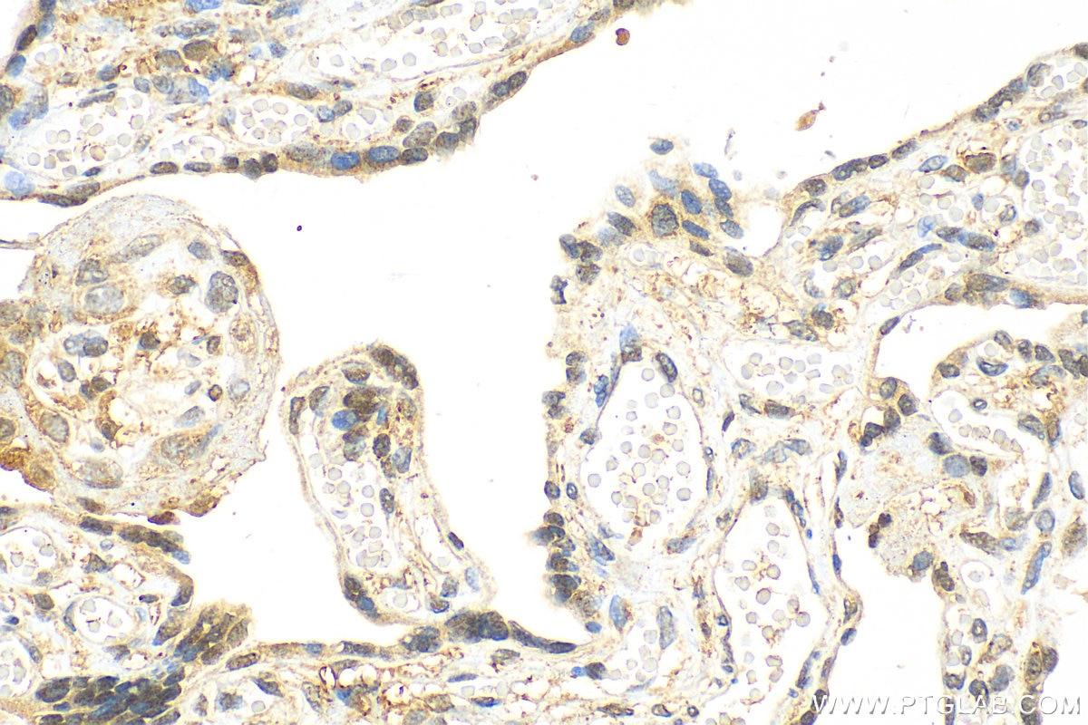
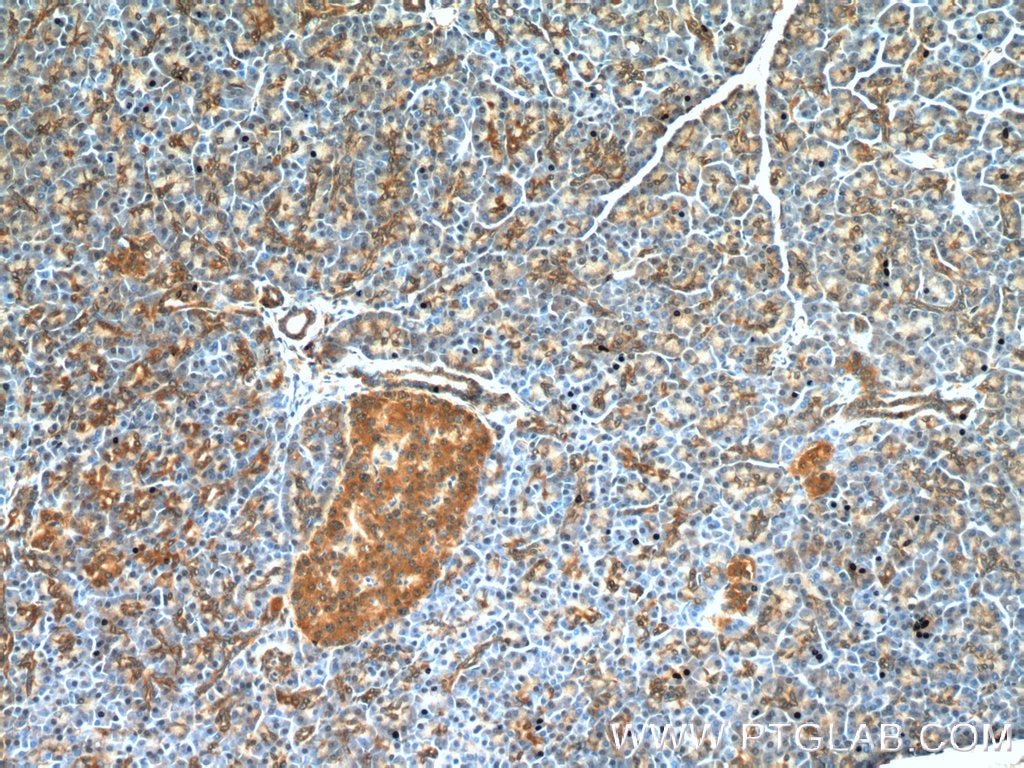
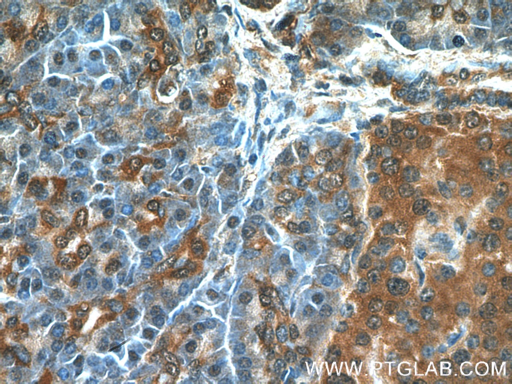
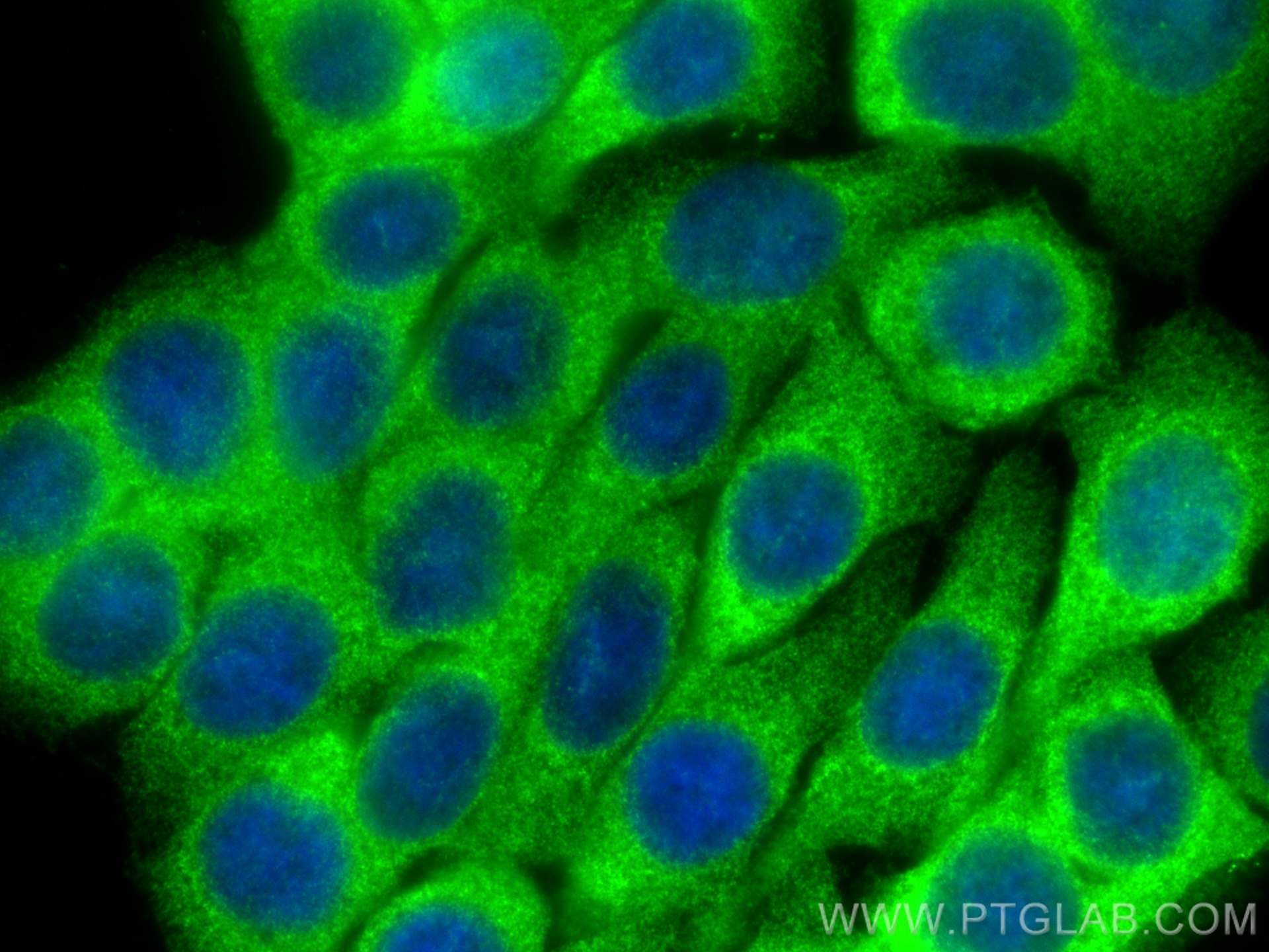
17563-1-PBS cible RIPK3 dans les applications de WB, IHC, IF/ICC, IP, Indirect ELISA et montre une réactivité avec des échantillons Humain
| Réactivité | Humain |
| Hôte / Isotype | Lapin / IgG |
| Clonalité | Polyclonal |
| Type | Anticorps |
| Immunogène | RIPK3 Protéine recombinante Ag11759 |
| Nom complet | receptor-interacting serine-threonine kinase 3 |
| Masse moléculaire calculée | 518 aa, 57 kDa |
| Poids moléculaire observé | 57-70 kDa |
| Numéro d’acquisition GenBank | BC062584 |
| Symbole du gène | RIPK3 |
| Identification du gène (NCBI) | 11035 |
| Conjugaison | Non conjugué |
| Forme | Liquide |
| Méthode de purification | Purification par affinité contre l'antigène |
| Tampon de stockage | PBS only |
| Conditions de stockage | Store at -80°C. 20ul contiennent 0,1% de BSA. |
Informations générales
Receptor-interacting protein 3 (RIP3, also known as RIPK3) is a serine-threonine protein involved in the regulation of inflammatory signaling and cell death. RIPK3, also named as RIP3, a Ser/Thr kinase of RIP (Receptor Interacting Protein) family, is a nucleocytoplasmic shuttling protein and its unconventional nuclear localization signal (NLS, 442-472 aa) is sufficient to trigger apoptosis in the nucleus (PMID: 18533105). It has 3 isoforms produced by alternative splicing.
What is the molecular weight of RIP3? Is RIP3 post-translationally modified?
The molecular weight of RIP3 is 57 kDa. During the induction of necroptosis, RIP3 migrates slower in SDS-PAGE due to its phosphorylation (PMID: 19524512). Additionally, RIP3 can be a subject of poly-ubiquitination, when targeted for degradation.
Are there any splice isoforms of RIP3?
Apart from full-length RIP3, there are two reported splice isoforms of RIP3: RIP3β and RIP3γ, and 28 and 25 kDa, respectively (PMID: 15896315).
What is the subcellular localization of RIP3?
RIP3 can shuttle between the cytoplasm and the nucleus. Although RIP3 forms necrosomes in the cytoplasm, a recent study suggests that the phosphorylation of RIP3, required for necroptosis, may also occur in the nucleus (PMID: 30271893). Additionally, the induction of necrosis by reactive oxygen species can cause transient translocation of RIP3 to mitochondria (PMID: 25206339).
What is the role of RIP3 in cell death (necroptosis)?
Activation of RIP3 kinase is required for the induction of necroptosis (PMID: 19524512, 19524513 and 19498109). Activation of RIP3 can be induced by interferons, death ligands, or by Toll-like receptors in response to pathogens. That leads to the phosphorylation of RIP3 and the formation of a β-amyloid-like protein complex. Phosphorylated RIP3 acts downstream by phosphorylation of MLKL (PMID: 30131615).
How to study necroptosis in a cell-based system?
The choice of the cell line is important. Many commonly used immortalized cell types are derived from cancers and may have very low RIL3 expression level (PMID: 25952668). Those cell lines are not going to be responsive to necrotic stimuli. A few of the examined cell types have high RIP3 levels: Jurkat, CCRF-CEM, U937, L929 cells, and mouse embryonic fibroblasts (PMID: 19524512). Good necroptosis readouts reflect an increased level of RIP3 protein and its phosphorylation.
