Anticorps Polyclonal de lapin anti-Syndecan-3
Syndecan-3 Polyclonal Antibody for WB, IHC, IF/ICC, IP, ELISA
Hôte / Isotype
Lapin / IgG
Réactivité testée
Humain, rat, souris
Applications
WB, IHC, IF/ICC, IP, ELISA
Conjugaison
Non conjugué
N° de cat : 10886-1-AP
Synonymes
Galerie de données de validation
 at dilution of 1:1000 incubated at room temperature for 1.5 hours.")
 at dilution of 1:500 incubated at room temperature for 1.5 hours.")
 at dilution of 1:500 incubated at room temperature for 1.5 hours.")
 at dilution of 1:500 incubated at room temperature for 1.5 hours.")
 at dilution of 1:500 incubated at room temperature for 1.5 hours.")
 at dilution of 1:500 incubated at room temperature for 1.5 hours.")
 with mouse lung tissue lysate 4000ug.")
 with A549 cells lysate 2800ug.")
. Heat mediated antigen retrieval with Tris-EDTA buffer (pH 9.0).")
. Heat mediated antigen retrieval with Tris-EDTA buffer (pH 9.0).")
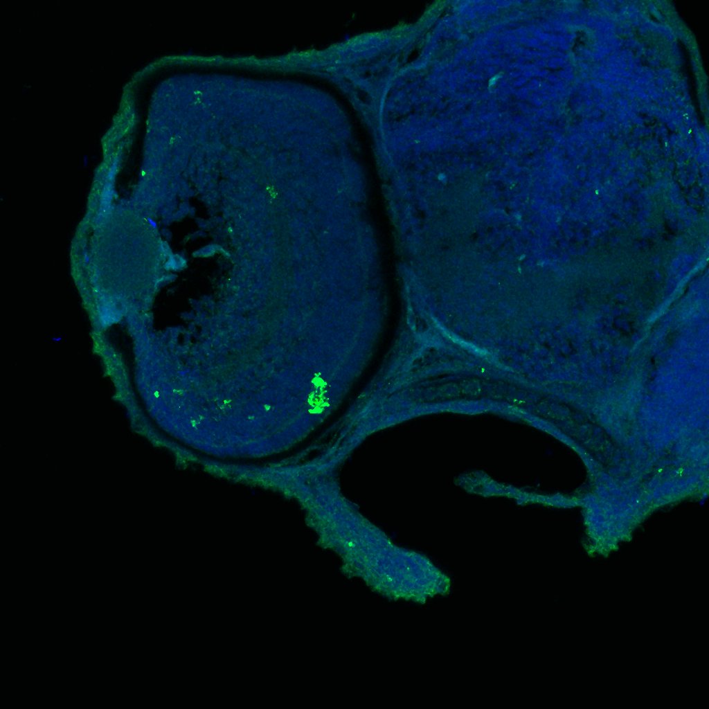
 fixed PC-12 cells using Syndecan-3 antibody (10886-1-AP) at dilution of 1:200 and CoraLite®488-Conjugated Goat Anti-Rabbit IgG(H+L).")
.")
Applications testées
| Résultats positifs en WB | cellules A549, cellules Jurkat, cellules L02, cellules MCF-7, cellules SH-SY5Y, tissu pulmonaire de rat, tissu pulmonaire de souris, tissu rénal de rat, tissu rénal de souris |
| Résultats positifs en IP | tissu pulmonaire de souris, cellules A549 |
| Résultats positifs en IHC | tissu de cancer du côlon humain il est suggéré de démasquer l'antigène avec un tampon de TE buffer pH 9.0; (*) À défaut, 'le démasquage de l'antigène peut être 'effectué avec un tampon citrate pH 6,0. |
| Résultats positifs en IF/ICC | cellules PC-12, cellules MCF-7 |
Dilution recommandée
| Application | Dilution |
|---|---|
| Western Blot (WB) | WB : 1:500-1:2000 |
| Immunoprécipitation (IP) | IP : 0.5-4.0 ug for 1.0-3.0 mg of total protein lysate |
| Immunohistochimie (IHC) | IHC : 1:50-1:500 |
| Immunofluorescence (IF)/ICC | IF/ICC : 1:50-1:500 |
| It is recommended that this reagent should be titrated in each testing system to obtain optimal results. | |
| Sample-dependent, check data in validation data gallery | |
Applications publiées
| WB | See 2 publications below |
| IHC | See 4 publications below |
| IF | See 1 publications below |
Informations sur le produit
10886-1-AP cible Syndecan-3 dans les applications de WB, IHC, IF/ICC, IP, ELISA et montre une réactivité avec des échantillons Humain, rat, souris
| Réactivité | Humain, rat, souris |
| Réactivité citée | Humain, souris |
| Hôte / Isotype | Lapin / IgG |
| Clonalité | Polyclonal |
| Type | Anticorps |
| Immunogène | Syndecan-3 Protéine recombinante Ag1317 |
| Nom complet | syndecan 3 |
| Masse moléculaire calculée | 38 kDa |
| Poids moléculaire observé | 60-70 kDa |
| Numéro d’acquisition GenBank | BC013974 |
| Symbole du gène | Syndecan-3 |
| Identification du gène (NCBI) | 9672 |
| Conjugaison | Non conjugué |
| Forme | Liquide |
| Méthode de purification | Purification par affinité contre l'antigène |
| Tampon de stockage | PBS with 0.02% sodium azide and 50% glycerol |
| Conditions de stockage | Stocker à -20°C. Stable pendant un an après l'expédition. L'aliquotage n'est pas nécessaire pour le stockage à -20oC Les 20ul contiennent 0,1% de BSA. |
Informations générales
Syndecan-3 is a member o f the Syndecan proteoglycan family. It plays a role in the organization of cell shape by affecting the actin cytoskeleton, possibly by transferring signals from the cell surface in a sugar-dependent mechanism.
Protocole
| Product Specific Protocols | |
|---|---|
| WB protocol for Syndecan-3 antibody 10886-1-AP | Download protocol |
| IHC protocol for Syndecan-3 antibody 10886-1-AP | Download protocol |
| IF protocol for Syndecan-3 antibody 10886-1-AP | Download protocol |
| IP protocol for Syndecan-3 antibody 10886-1-AP | Download protocol |
| Standard Protocols | |
|---|---|
| Click here to view our Standard Protocols |
Publications
| Species | Application | Title |
|---|---|---|
Nucleic Acids Res Metabolic and chemical regulation of tRNA modification associated with taurine deficiency and human disease. | ||
Am J Pathol No haploinsufficiency but loss of heterozygosity for EXT in multiple osteochondromas. | ||
Am J Pathol Screening for potential targets for therapy in mesenchymal, clear cell, and dedifferentiated chondrosarcoma reveals Bcl-2 family members and TGFβ as potential targets. | ||
J Cell Sci Electrophoresis of cell membrane heparan sulfate regulates galvanotaxis in glial cells. | ||
BMC Cancer Prognostic significance of the expression of GFRα1, GFRα3 and syndecan-3, proteins binding ARTEMIN, in mammary carcinoma. | ||
Medicine (Baltimore) Two novel predictive biomarkers for osteosarcoma and glycolysis pathways: A profiling study on HS2ST1 and SDC3 |
Avis
The reviews below have been submitted by verified Proteintech customers who received an incentive for providing their feedback.
FH Kamal (Verified Customer) (02-15-2024) | Mouse liver lysates were subjected to SDS PAGE followed by western blot with 10886-1-AP (Syndecan-3 antibody) at dilution of 1:2000 in 1X TBST incubated overnight at 4 degree C. Syndecan-3 appeared at 65 kDa.
|
FH Ryan (Verified Customer) (02-28-2019) | Tissue was fixed in PFA with tris-HCl antigen retrieval ph=6. Co-localisation with microglia based on known markers (not shown).
 |

