- Phare
- Validé par KD/KO
Anticorps Polyclonal de lapin anti-GLUT1
GLUT1 Polyclonal Antibody for WB, IHC, IF/ICC, IF-P, FC (Intra), ELISA
Hôte / Isotype
Lapin / IgG
Réactivité testée
Humain, rat, souris et plus (4)
Applications
WB, IHC, IF/ICC, IF-P, FC (Intra), ChIP, ELISA
Conjugaison
Non conjugué
N° de cat : 21829-1-AP
Synonymes
Galerie de données de validation
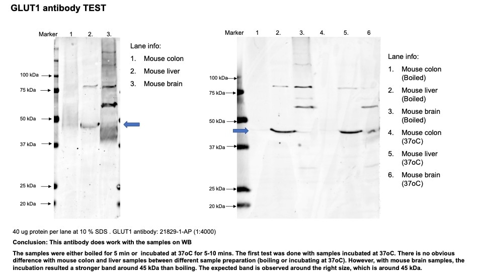
 were subjected to SDS PAGE followed by western blot with 21829-1-AP (GLUT1 antibody) at dilution of 1:4000 incubated at room temperature for 1.5 hours.")
 were subjected to SDS PAGE followed by western blot with 21829-1-AP (GLUT1 antibody) at dilution of 1:4000 incubated at room temperature for 1.5 hours.")
 were subjected to SDS PAGE followed by western blot with 21829-1-AP (GLUT1 antibody) at dilution of 1:4000 incubated at room temperature for 1.5 hours.")
 at dilution of 1:1000 (under 10x lens). Heat mediated antigen retrieval with Tris-EDTA buffer (pH 9.0).")
 at dilution of 1:5000 (under 40x lens). Heat mediated antigen retrieval with Tris-EDTA buffer (pH 9.0).")
 at dilution of 1:1000 (under 10x lens). Heat mediated antigen retrieval with Tris-EDTA buffer (pH 9.0).")
 at dilution of 1:1000 (under 10x lens). Heat mediated antigen retrieval with Tris-EDTA buffer (pH 9.0).")
 fixed mouse brain tissue using GLUT1 antibody (21829-1-AP) at dilution of 1:2000 and CoraLite®488-Conjugated AffiniPure Goat Anti-Rabbit IgG(H+L).")
 fixed HeLa cells using GLUT1 antibody (21829-1-AP) at dilution of 1:400 and CoraLite®488-Conjugated AffiniPure Goat Anti-Rabbit IgG(H+L).")
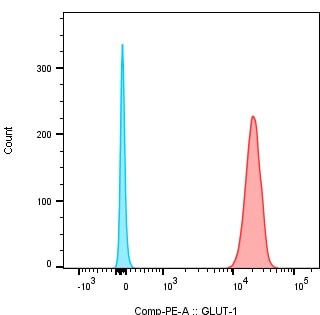
(red), or 0.4 ug rabbit IgG isotype control (blue). Cells were fixed with 4% PFA and permeabilized with Flow Cytometry Perm Buffer (PF00011-C).")
Applications testées
| Résultats positifs en WB | unboiled HT-29 cells, tissu de côlon de souris incubé à 37 °C |
| Résultats positifs en IHC | tissu cérébral de rat, tissu de cancer du col de l'utérus humain, tissu de cancer du poumon humain, tissu de cancer du sein humain il est suggéré de démasquer l'antigène avec un tampon de TE buffer pH 9.0; (*) À défaut, 'le démasquage de l'antigène peut être 'effectué avec un tampon citrate pH 6,0. |
| Résultats positifs en IF-P | tissu cérébral de souris, |
| Résultats positifs en IF/ICC | cellules HeLa, |
| Résultats positifs en FC (Intra) | cellules HeLa |
Dilution recommandée
| Application | Dilution |
|---|---|
| Western Blot (WB) | WB : 1:1000-1:8000 |
| Immunohistochimie (IHC) | IHC : 1:2500-1:10000 |
| Immunofluorescence (IF)-P | IF-P : 1:1000-1:4000 |
| Immunofluorescence (IF)/ICC | IF/ICC : 1:200-1:800 |
| Flow Cytometry (FC) (INTRA) | FC (INTRA) : 0.40 ug per 10^6 cells in a 100 µl suspension |
| It is recommended that this reagent should be titrated in each testing system to obtain optimal results. | |
| Sample-dependent, check data in validation data gallery | |
Applications publiées
| KD/KO | See 7 publications below |
| WB | See 274 publications below |
| IHC | See 53 publications below |
| IF | See 53 publications below |
| ChIP | See 1 publications below |
Informations sur le produit
21829-1-AP cible GLUT1 dans les applications de WB, IHC, IF/ICC, IF-P, FC (Intra), ChIP, ELISA et montre une réactivité avec des échantillons Humain, rat, souris
| Réactivité | Humain, rat, souris |
| Réactivité citée | rat, Chèvre, Humain, Lapin, porc, souris, Lasiopodomys Brandtii |
| Hôte / Isotype | Lapin / IgG |
| Clonalité | Polyclonal |
| Type | Anticorps |
| Immunogène | GLUT1 Protéine recombinante Ag16282 |
| Nom complet | solute carrier family 2 (facilitated glucose transporter), member 1 |
| Masse moléculaire calculée | 492 aa, 54 kDa |
| Poids moléculaire observé | 45-55 kDa |
| Numéro d’acquisition GenBank | BC121804 |
| Symbole du gène | GLUT1 |
| Identification du gène (NCBI) | 6513 |
| Conjugaison | Non conjugué |
| Forme | Liquide |
| Méthode de purification | Purification par affinité contre l'antigène |
| Tampon de stockage | PBS with 0.02% sodium azide and 50% glycerol |
| Conditions de stockage | Stocker à -20°C. Stable pendant un an après l'expédition. L'aliquotage n'est pas nécessaire pour le stockage à -20oC Les 20ul contiennent 0,1% de BSA. |
Informations générales
Glucose transporter 1 (GLUT1), also known as solute carrier family 2, facilitated glucose transporter member 1 (SLC2A1), is a uniporter protein responsible for the transport of glucose in many cell types and across the blood-brain barrier.
What is the molecular weight of GLUT1? Is GLUT1 post-translationally modified?
There are two forms of GLUT1 transporter that differ in their molecular weight. The 45-kDa form is found in glial cells, while the 55-kDa form is present in the endothelial cells regulating glucose transport over the blood-brain and blood-tissue barriers (PMID: 9630522). N-glycosylation of asparagine at position 42 is the only known post-translation modification of GLUT1 (PMID: 3839598).
What is the subcellular localization of GLUT1?
Glucose transporters, including GLUT1, are multiple-pass integral membrane proteins. GLUT1 is present at the plasma membrane but is also a subject of recycling between plasma membrane and endosomes.
What molecules can be transported by GLUT1?
The main substrate of GLUT1 transport is glucose, but it can also transport galactose, mannose, glucosamine, and reduced ascorbate.
What is the tissue expression pattern of GLUT1?
GLUT1 is expressed by many cell types but the highest levels are observed in erythrocytes and in the central nervous system (astrocytes). GLUT1 is responsible for glucose transfer across the blood-brain and blood-tissue barriers, including placental transport.
Protocole
| Product Specific Protocols | |
|---|---|
| WB protocol for GLUT1 antibody 21829-1-AP | Download protocol |
| IHC protocol for GLUT1 antibody 21829-1-AP | Download protocol |
| IF protocol for GLUT1 antibody 21829-1-AP | Download protocol |
| Standard Protocols | |
|---|---|
| Click here to view our Standard Protocols |
Publications
| Species | Application | Title |
|---|---|---|
Adv Mater Supramolecular Hydrogel with Ultra-Rapid Cell-Mediated Network Adaptation for Enhancing Cellular Metabolic Energetics and Tissue Regeneration | ||
Int J Oral Sci Transcriptional activation of glucose transporter 1 in orthodontic tooth movement-associated mechanical response. | ||
ACS Nano Biomimetic Nanomedicine Targeting Orchestrated Metabolism Coupled with Regulatory Factors to Disrupt the Metabolic Plasticity of Breast Cancer | ||
Nat Commun Parvimonas micra promotes oral squamous cell carcinoma metastasis through TmpC-CKAP4 axis | ||
Neuron Sympathetic nerve-enteroendocrine L cell communication modulates GLP-1 release, brain glucose utilization, and cognitive function |
Avis
The reviews below have been submitted by verified Proteintech customers who received an incentive for providing their feedback.
FH Marco (Verified Customer) (07-04-2024) | Nice Westernblots in NRVCMs
|
FH Hua (Verified Customer) (02-14-2023) | Good antibody working for WB with mouse liver samples. However, boiling mouse colon and brain tissues results in no band observed.
 |
FH William (Verified Customer) (10-26-2021) | Very clear staining in IHC in rat brain tissue (Wistar) at a concentration of 1:100, very well localised to blood vessels. Not tested yet at different concentrations
|
FH Bastien (Verified Customer) (08-19-2020) | Staining of B cells from mice bone marrow after cytoplasmic permezbiliation. The cells were first fixed and permeabilized with intracellular fix and perm set from ebioscience and then stained with 1 µl/ million cells with Glut-1 antibody (21829-1-AP) during 50min After, a seconde staining was performed with 0,1µl/million cells of F(ab')2-Donkey anti-Rabbit IgG (H+L), PE, Secondary Antibody from invitrogen. In blue secondary antibody alone and in red primary (21829-1-AP) +secondary antibody
 |
FH Susan (Verified Customer) (11-19-2019) | 20ug of HeLa cells overexpressing Glut1-GFP. Blocked with 5% milk in 0.1% TBST and incubated overnight at 4 degrees with rocking.
 |
FH Kishor (Verified Customer) (12-13-2018) | I got good results in cultured cells but I could not see any results with rat liver protein.
|

