- Phare
- Validé par KD/KO
Anticorps Polyclonal de lapin anti-P62/SQSTM1
P62/SQSTM1 Polyclonal Antibody for WB, IHC, IF/ICC, FC (Intra), IP, ELISA
Hôte / Isotype
Lapin / IgG
Réactivité testée
Humain et plus (6)
Applications
WB, IHC, IF/ICC, FC (Intra), IP, CoIP, ELISA
Conjugaison
Non conjugué
N° de cat : 18420-1-AP
Synonymes
Galerie de données de validation
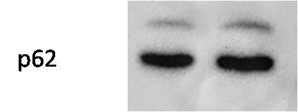
 at dilution of 1:10000 incubated at room temperature for 1.5 hours.")
 at dilution of 1:4000 incubated at room temperature for 1.5 hours.")
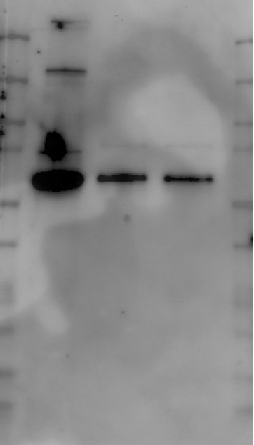
 lysates prepared with RIPA buffer, 25 μg protein loaded. 18420-1-AP incubated at 1:1000 at 4°C overnight in 5% milk in TBST. Ponceau stained transfers shown on right. Data provided by YCharOS, an open science company with a mission to validate commercial antibodies to improve scientific reproducibility and transparency.")
 with sh-Control and sh-P62/SQSTM1 transfected HEK-293 cells.")

 with HepG2 cells lysate 1360 ug.")
 at dilution of 1:50 (under 40x lens).")
 at dilution of 1:200 (under 10x lens). Heat mediated antigen retrieval with Tris-EDTA buffer (pH 9.0).")
 at dilution of 1:200 (under 40x lens). Heat mediated antigen retrieval with Tris-EDTA buffer (pH 9.0).")
 at dilution of 1:400 (under 10x lens). Heat mediated antigen retrieval with Tris-EDTA buffer (pH 9.0).")
 at dilution of 1:400 (under 40x lens). Heat mediated antigen retrieval with Tris-EDTA buffer (pH 9.0).")
 at dilution of 1:400 (under 10x lens). Heat mediated antigen retrieval with Tris-EDTA buffer (pH 9.0).")
 at dilution of 1:400 (under 40x lens). Heat mediated antigen retrieval with Tris-EDTA buffer (pH 9.0).")
.")
 fixed Chloroquine treated HepG2 cells using P62,SQSTM1 antibody (18420-1-AP) at dilution of 1:1500 and CoraLite®488-Conjugated AffiniPure Goat Anti-Rabbit IgG(H+L), CL594-Phalloidin (red).")
 fixed Chloroquine treated HeLa cells using P62,SQSTM1 antibody (18420-1-AP) at dilution of 1:500 and CoraLite®488-Conjugated AffiniPure Goat Anti-Rabbit IgG(H+L), (CL594-Phalloidin, red).")
 and SQSTM1 KO cells (red outline) labelled with a green or a far-red fluorescence dye, respectively. Cells fixed with 4% PFA and stained with 18420-1-AP at 1:300. Bars = 10 μm. Data provided by YCharOS, an open science company with a mission to validate commercial antibodies to improve scientific reproducibility and transparency.")
 and CoraLite®488-Conjugated AffiniPure Goat Anti-Rabbit IgG(H+L) at dilution 1:1000 (red), or 0.4 ug Isotype Control. Cells were fixed with 4% PFA and permeabilized with Flow Cytometry Perm Buffer (PF00011-C).")
Applications testées
| Résultats positifs en WB | cellules HeLa, cellules HEK-293, cellules HepG2, cellules Jurkat, cellules MG U-87, cellules U2OS |
| Résultats positifs en IP | cellules HepG2, cellules U2OS |
| Résultats positifs en IHC | tissu de cancer du poumon humain, tissu de cancer du foie humain, tissu de gliome humain il est suggéré de démasquer l'antigène avec un tampon de TE buffer pH 9.0; (*) À défaut, 'le démasquage de l'antigène peut être 'effectué avec un tampon citrate pH 6,0. |
| Résultats positifs en IF/ICC | cellules HepG2 traitées à la chloroquine, cellules HeLa traitées à la chloroquine, cellules U2OS |
| Résultats positifs en FC (Intra) | cellules HEK-293 |
Dilution recommandée
| Application | Dilution |
|---|---|
| Western Blot (WB) | WB : 1:5000-1:50000 |
| Immunoprécipitation (IP) | IP : 0.5-4.0 ug for 1.0-3.0 mg of total protein lysate |
| Immunohistochimie (IHC) | IHC : 1:200-1:800 |
| Immunofluorescence (IF)/ICC | IF/ICC : 1:750-1:3000 |
| Flow Cytometry (FC) (INTRA) | FC (INTRA) : 0.40 ug per 10^6 cells in a 100 µl suspension |
| It is recommended that this reagent should be titrated in each testing system to obtain optimal results. | |
| Sample-dependent, check data in validation data gallery | |
Applications publiées
| KD/KO | See 41 publications below |
| WB | See 1731 publications below |
| IHC | See 168 publications below |
| IF | See 218 publications below |
| IP | See 23 publications below |
| CoIP | See 13 publications below |
Informations sur le produit
18420-1-AP cible P62/SQSTM1 dans les applications de WB, IHC, IF/ICC, FC (Intra), IP, CoIP, ELISA et montre une réactivité avec des échantillons Humain
| Réactivité | Humain |
| Réactivité citée | Chèvre, Humain, Lapin, poisson-zèbre, poulet, singe, oisons |
| Hôte / Isotype | Lapin / IgG |
| Clonalité | Polyclonal |
| Type | Anticorps |
| Immunogène | P62/SQSTM1 Protéine recombinante Ag13131 |
| Nom complet | sequestosome 1 |
| Masse moléculaire calculée | 48 kDa |
| Poids moléculaire observé | 62 kDa |
| Numéro d’acquisition GenBank | BC017222 |
| Symbole du gène | P62/SQSTM1 |
| Identification du gène (NCBI) | 8878 |
| Conjugaison | Non conjugué |
| Forme | Liquide |
| Méthode de purification | Purification par affinité contre l'antigène |
| Tampon de stockage | PBS with 0.02% sodium azide and 50% glycerol |
| Conditions de stockage | Stocker à -20°C. Stable pendant un an après l'expédition. L'aliquotage n'est pas nécessaire pour le stockage à -20oC Les 20ul contiennent 0,1% de BSA. |
Informations générales
Background
P62 (ubiquitin-binding protein P62), also known as Sequestosome-1, is a multifunctional adaptor protein most widely known for its role as an autophagosome cargo protein (PMID: 8551575). P62 via specific interactions with polyubiquitylated target proteins induces their selective autophagy (PMID: 17580304). It also plays an important role in the regulation of the NFkB signaling pathway, senescence, cell differentiation, apoptosis, and immune responses (PMID: 26404812).
What is the molecular weight of P62?
The observed molecular weight of the protein can vary from as low as 8 kDa (for the smallest isoforms) to 48 kDa.
What is the subcellular localization of P62?
P62 is mainly localized in the cytoplasm; however, upon autophagy induction, e.g., via starvation or selective inhibitor treatment, it localizes in vesicular structures - autophagosomes.
What is the tissue specificity of P62?
It is ubiquitously expressed in various tissues.
What is the function of P62 in the regulation of cell death and autophagy?
It is a selective autophagy receptor that forms a bridge between polyubiquitylated cargo (via its UBA domains) and an autophagy modifier such as LC3 (via LIR domains) (PMIDs: 16286508, 20168092, 24128730, 28404643, 22622177). The process of selective autophagy is tightly regulated at many levels, including the posttranslational modifications (PTMs) of various proteins in the cascade, P62 among others (PMID: 29233872). P62 is involved in the regulation of cell death induction in response to various stimuli, e.g., via activation of caspase-8 at the autophagosome membrane (PMID: 29480462). In addition, P62 is degraded during the autophagic process, which makes its intracellular level a marker for autophagy progression.
What is P62's involvement in disease?
Mutations in P62 have been associated with the following diseases: sporadic and familial Paget's disease of bone, neurodegenerative diseases, diabetes, and obesity (PMID: 29480462). A growing number of reports suggest the implication of P62 in the induction of multiple cellular oncogenic transformations. Indeed, increased levels of P62 have been linked to tumor formation, cancer promotion, and resistance to therapy (PMID: 29738493). Moreover, P62 is an unfavorable prognostic marker in liver cancer.
Protocole
| Product Specific Protocols | |
|---|---|
| WB protocol for P62/SQSTM1 antibody 18420-1-AP | Download protocol |
| IHC protocol for P62/SQSTM1 antibody 18420-1-AP | Download protocol |
| IF protocol for P62/SQSTM1 antibody 18420-1-AP | Download protocol |
| IP protocol for P62/SQSTM1 antibody 18420-1-AP | Download protocol |
| Standard Protocols | |
|---|---|
| Click here to view our Standard Protocols |
Publications
| Species | Application | Title |
|---|---|---|
Nature FBXO38 mediates PD-1 ubiquitination and regulates anti-tumour immunity of T cells. | ||
Signal Transduct Target Ther Targeting CRL4 suppresses chemoresistant ovarian cancer growth by inducing mitophagy | ||
Mol Cancer Overcoming multi-drug resistance in SCLC: a synergistic approach with venetoclax and hydroxychloroquine targeting the lncRNA LYPLAL1-DT/BCL2/BECN1 pathway | ||
Cell Metab Disrupted methionine cycle triggers muscle atrophy in cancer cachexia through epigenetic regulation of REDD1 | ||
Cancer Cell iASPP Is an Antioxidative Factor and Drives Cancer Growth and Drug Resistance by Competing with Nrf2 for Keap1 Binding. | ||
Cell Metab Receptor-Mediated ER Export of Lipoproteins Controls Lipid Homeostasis in Mice and Humans. |
Avis
The reviews below have been submitted by verified Proteintech customers who received an incentive for providing their feedback.
FH David (Verified Customer) (01-02-2024) | Used for WB in cell lysates. Prominent band at the predicted molecular weight, although some non-specific bands in the control at around 40kDa. However, the correct SQSTM1 band can be clearly visualised.
|
FH Alexandru (Verified Customer) (11-21-2023) | Great at detecting p62 in cell lysates.
|
FH Wan (Verified Customer) (02-24-2022) | Perfect antibody, clear bands!
|
FH X (Verified Customer) (01-18-2021) | It is a reliable Ab to detect p62 with mouse brain lysate, although there is other band detected.
 |
FH X (Verified Customer) (01-18-2021) | It is a reliable Ab to detect p62 with mouse brain lysate in WB, although there is other bands seen.
|
FH Wei (Verified Customer) (01-30-2020) | Good, Clear band for mouse heart
|
FH Joleen (Verified Customer) (06-16-2019) | Recognizes SQSTM1 prominently with some non-specific bands.
 |

