- Phare
- Validé par KD/KO
Anticorps Polyclonal de lapin anti-TBC1D5
TBC1D5 Polyclonal Antibody for WB, IHC, IP, ELISA
Hôte / Isotype
Lapin / IgG
Réactivité testée
Humain, souris
Applications
WB, IHC, IF, IP, ELISA
Conjugaison
Non conjugué
N° de cat : 17078-1-AP
Synonymes
Galerie de données de validation
 at dilution of 1:5000 incubated at room temperature for 1.5 hours.")
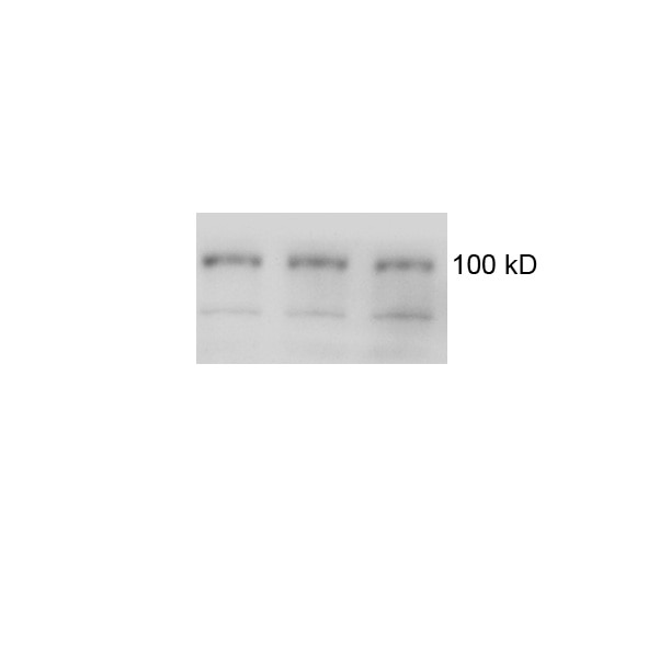
 with Jurkat cells lysate 1360 ug.")
 at dilution of 1:200 (under 40x lens). Heat mediated antigen retrieval with Tris-EDTA buffer (pH 9.0).")
 at dilution of 1:200 (under 10x lens). Heat mediated antigen retrieval with Tris-EDTA buffer (pH 9.0).")
Applications testées
| Résultats positifs en WB | cellules A431, cellules HEK-293T, cellules Jurkat, cellules PC-3 |
| Résultats positifs en IP | cellules Jurkat, |
| Résultats positifs en IHC | tissu testiculaire de souris, il est suggéré de démasquer l'antigène avec un tampon de TE buffer pH 9.0; (*) À défaut, 'le démasquage de l'antigène peut être 'effectué avec un tampon citrate pH 6,0. |
Dilution recommandée
| Application | Dilution |
|---|---|
| Western Blot (WB) | WB : 1:2000-1:10000 |
| Immunoprécipitation (IP) | IP : 0.5-4.0 ug for 1.0-3.0 mg of total protein lysate |
| Immunohistochimie (IHC) | IHC : 1:50-1:500 |
| It is recommended that this reagent should be titrated in each testing system to obtain optimal results. | |
| Sample-dependent, check data in validation data gallery | |
Applications publiées
| KD/KO | See 5 publications below |
| WB | See 13 publications below |
| IF | See 6 publications below |
Informations sur le produit
17078-1-AP cible TBC1D5 dans les applications de WB, IHC, IF, IP, ELISA et montre une réactivité avec des échantillons Humain, souris
| Réactivité | Humain, souris |
| Réactivité citée | Humain, souris |
| Hôte / Isotype | Lapin / IgG |
| Clonalité | Polyclonal |
| Type | Anticorps |
| Immunogène | TBC1D5 Protéine recombinante Ag10991 |
| Nom complet | TBC1 domain family, member 5 |
| Masse moléculaire calculée | 795 aa, 89 kDa |
| Poids moléculaire observé | 89 kDa |
| Numéro d’acquisition GenBank | BC013145 |
| Symbole du gène | TBC1D5 |
| Identification du gène (NCBI) | 9779 |
| Conjugaison | Non conjugué |
| Forme | Liquide |
| Méthode de purification | Purification par affinité contre l'antigène |
| Tampon de stockage | PBS with 0.02% sodium azide and 50% glycerol |
| Conditions de stockage | Stocker à -20°C. Stable pendant un an après l'expédition. L'aliquotage n'est pas nécessaire pour le stockage à -20oC Les 20ul contiennent 0,1% de BSA. |
Informations générales
TBC1D5, a member of TBC (Tre2/Bub2/Cdc16)1 domain family, is a novel retromer-interacting protein. TBC1D5 is a Rab GTPase-activating proteins (GAPs) proteins that negatively regulates VPS35/29/26 recruitment and causes Rab7 to dissociate from the membrane. TBC1D5 could bridge the endosome and autophagosome via its C-terminal LIR motif, and Rab GAPs are implicated in the reprogramming of the endocytic trafficking events under starvation-induced autophagy. The TBC1D5 protein exists 89 kDa and 91 kDa isoforms.
Protocole
| Product Specific Protocols | |
|---|---|
| WB protocol for TBC1D5 antibody 17078-1-AP | Download protocol |
| IHC protocol for TBC1D5 antibody 17078-1-AP | Download protocol |
| IP protocol for TBC1D5 antibody 17078-1-AP | Download protocol |
| Standard Protocols | |
|---|---|
| Click here to view our Standard Protocols |
Publications
| Species | Application | Title |
|---|---|---|
Sci Adv De novo macrocyclic peptides for inhibiting, stabilizing, and probing the function of the retromer endosomal trafficking complex. | ||
EMBO J Control of RAB7 activity and localization through the retromer-TBC1D5 complex enables RAB7-dependent mitophagy.
| ||
J Cell Biol Retromer and TBC1D5 maintain late endosomal RAB7 domains to enable amino acid-induced mTORC1 signaling.
| ||
EMBO Rep BioID reveals an ATG9A interaction with ATG13-ATG101 in the degradation of p62/SQSTM1-ubiquitin clusters. | ||
Int J Biol Sci TBK1 Facilitates GLUT1-Dependent Glucose Consumption by suppressing mTORC1 Signaling in Colorectal Cancer Progression.
| ||
Mol Biol Cell Trafficking defects in WASH-knockout fibroblasts originate from collapsed endosomal and lysosomal networks. |
Avis
The reviews below have been submitted by verified Proteintech customers who received an incentive for providing their feedback.
FH q (Verified Customer) (06-01-2021) | It is OK to detect TBC1D5 (100 kD) in both mouse and postmortem brain lysates, although there are some non specific bands.
 |
FH Florian (Verified Customer) (04-09-2019) | This is a very strong antibody that is reasonably specific in western blot applications. We have validated it through Crispr/Cas9 mediated knockout of TBC1D5 in HeLa cells (Jimenez-Orgaz et al, EMBOJ, 2018). While the strongest band is indeed TBC1D5, as our knockout confirms, it also produces several weaker, unspecific bands on membranes blocked with 5% milk. Because of the unspecific bands, I rate it with four stars instead of five. We have not tested it for immunofluorescence. Overall, it is a very good antibody that will probably also work at much lower dilutions than 1:1000. Diluting it further may also reduce the unspecific signal.
|
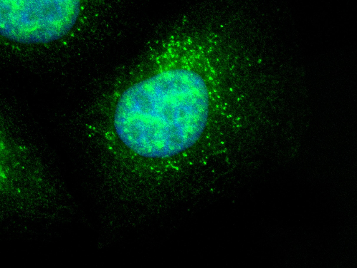
FH David (Verified Customer) (02-28-2019) | Hela cells stained with anti-TBC1D5 (green) and Hoechst (blue)
 |

