- Phare
- Validé par KD/KO
Anticorps Monoclonal anti-TDP-43 (human specific)
TDP-43 (human specific) Monoclonal Antibody for WB, IHC, IP, Sandwich ELISA, Indirect ELISA, Sample test
Hôte / Isotype
Mouse / IgG1
Réactivité testée
Humain
Applications
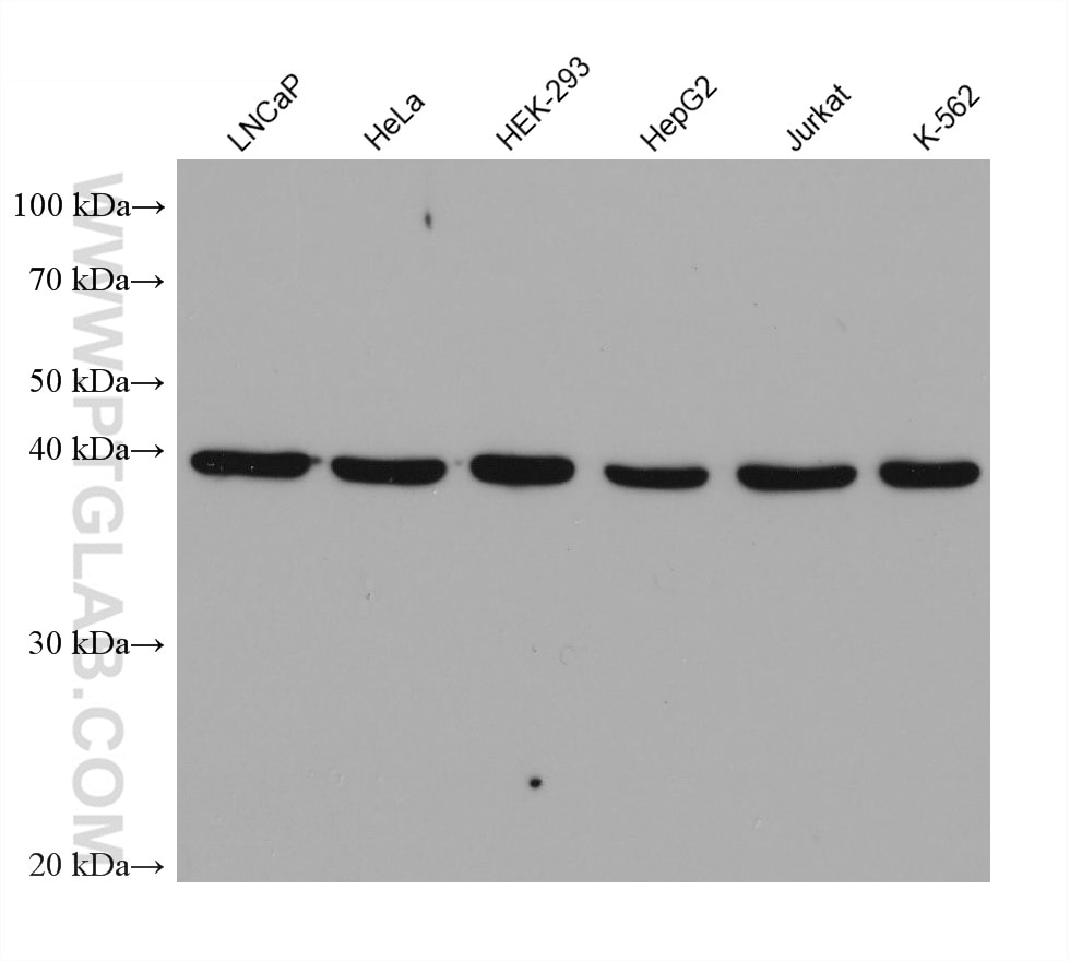
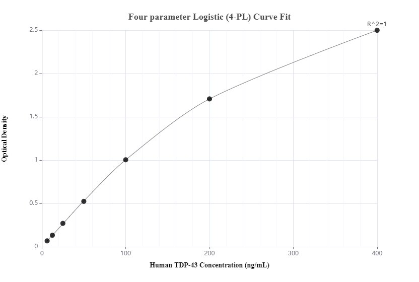
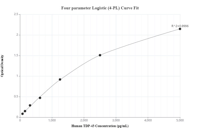
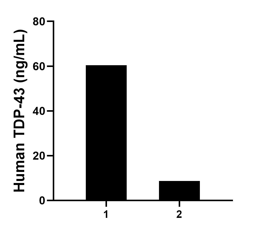
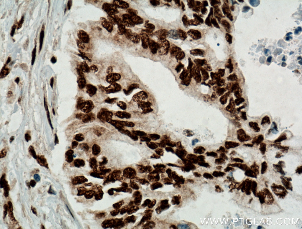
WB, IHC, IP, Sandwich ELISA, Indirect ELISA, Sample test
Conjugaison
Non conjugué
CloneNo.
6H6E12
N° de cat : 60019-2-PBS
Synonymes
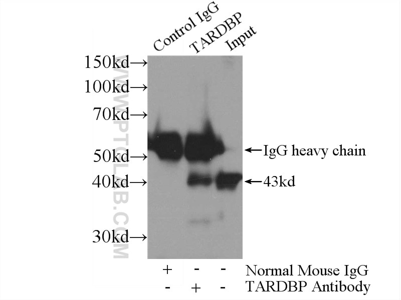
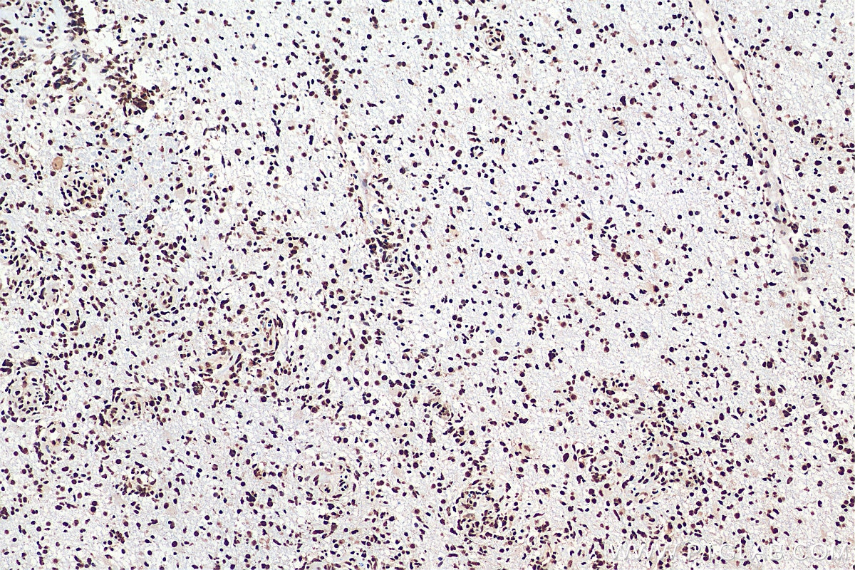
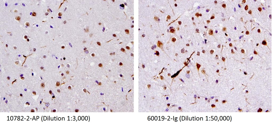
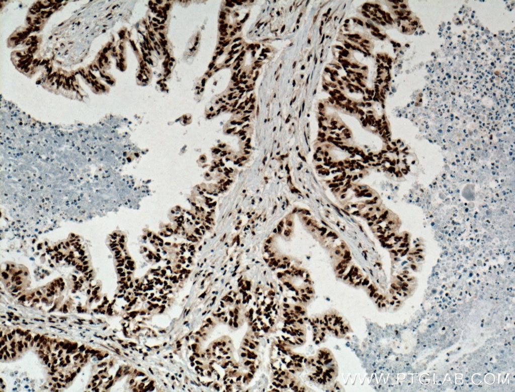
Galerie de données de validation
Informations sur le produit
60019-2-PBS cible TDP-43 (human specific) dans les applications de WB, IHC, IP, Sandwich ELISA, Indirect ELISA, Sample test et montre une réactivité avec des échantillons Humain
| Réactivité | Humain |
| Hôte / Isotype | Mouse / IgG1 |
| Clonalité | Monoclonal |
| Type | Anticorps |
| Immunogène | Protéine recombinante |
| Nom complet | TAR DNA binding protein |
| Masse moléculaire calculée | 43 kDa |
| Poids moléculaire observé | 43 kDa |
| Numéro d’acquisition GenBank | BC001487 |
| Symbole du gène | TDP-43 |
| Identification du gène (NCBI) | 23435 |
| Conjugaison | Non conjugué |
| Forme | Liquide |
| Méthode de purification | Purification par protéine G |
| Tampon de stockage | PBS only |
| Conditions de stockage | Store at -80°C. 20ul contiennent 0,1% de BSA. |
Informations générales
Transactivation response (TAR) DNA-binding protein of 43 kDa (also known as TARDBP or TDP-43) was first isolated as a transcriptional inactivator binding to the TAR DNA element of the HIV-1 virus. Neumann et al. (2006) found that a hyperphosphorylated, ubiquitinated, and cleaved form of TARDBP, known as pathologic TDP-43, is the major component of the tau-negative and ubiquitin-positive inclusions that characterize amyotrophic lateral sclerosis (ALS) and the most common pathological subtype of frontotemporal lobar degeneration (FTLD-U). Various forms of TDP-43 exist, including 18-35 kDa of cleaved C-terminal fragments, 45-50 kDa phospho-protein, 55 kDa glycosylated form, 75 kDa hyperphosphorylated form, and 90-300 kDa cross-linked form. (PMID: 17023659,19823856, 21666678, 22193176). 60019-2-Ig is a mouse monoclonal antibody recognizing the cleavage product of 20-30 kDa in addition to the native and phosphorylated forms of TDP-43. Immunohistochemical analyses of TDP-43 using this antibody detect both normal diffuse nuclear staining and insoluble inclusions in pathologic tissues. Notably this antibody only recognizes human TDP-43 but not reacts with mouse or rat TDP-43.
