- Phare
- Validé par KD/KO
Anticorps Polyclonal de lapin anti-TIA1
TIA1 Polyclonal Antibody for WB, IHC, IF/ICC, FC (Intra), IP, ELISA
Hôte / Isotype
Lapin / IgG
Réactivité testée
Humain, rat, souris et plus (2)
Applications
WB, IHC, IF/ICC, FC (Intra), IP, CoIP, ChIP, RIP, ELISA
Conjugaison
Non conjugué
N° de cat : 12133-2-AP
Synonymes
Galerie de données de validation
 at dilution of 1:1500 incubated at room temperature for 1.5 hours.")
 at dilution of 1:40000 incubated at room temperature for 1.5 hours.")
 at dilution of 1:20000 incubated at room temperature for 1.5 hours.")
 with sh-Control and sh-TIA1 transfected Jurkat cells.")
 at dilution of 1:1500 incubated at room temperature for 1.5 hours.")
 at dilution of 1:2000 incubated at room temperature for 1.5 hours.")
 at dilution of 1:1000 incubated at room temperature for 1.5 hours.")
 at dilution of 1:1000 incubated at room temperature for 1.5 hours.")
 with Jurkat cells lysate 3200ug.")
 at dilution of 1:200 (under 40x lens). Heat mediated antigen retrieval with Tris-EDTA buffer (pH 9.0).")
 at dilution of 1:200 (under 10x lens). Heat mediated antigen retrieval with Tris-EDTA buffer (pH 9.0).")
 at dilution of 1:200 (under 40x lens). Heat mediated antigen retrieval with Tris-EDTA buffer (pH 9.0).")
 at dilution of 1:200 (under 10x lens). Heat mediated antigen retrieval with Tris-EDTA buffer (pH 9.0).")
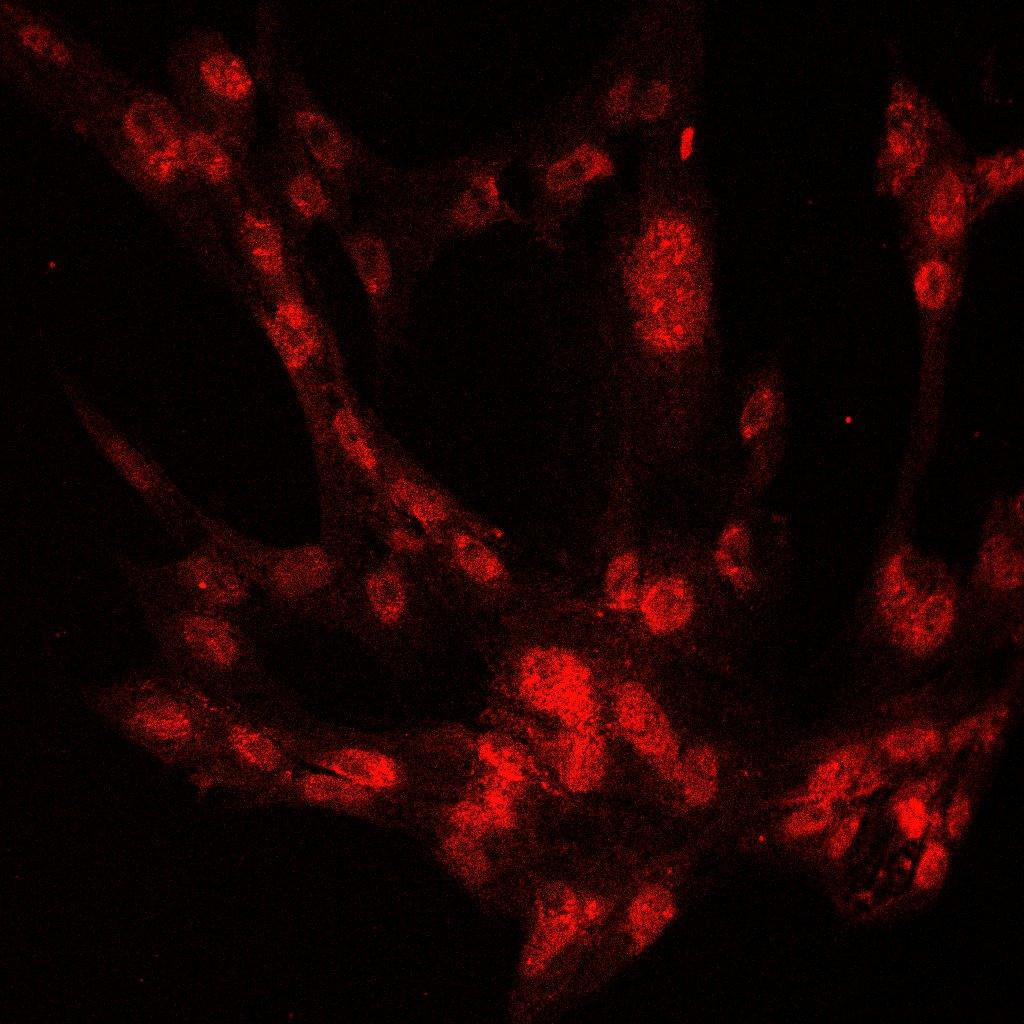
 fixed L02 cells using TIA1 antibody (12133-2-AP) at dilution of 1:200 and CoraLite®488-Conjugated Goat Anti-Rabbit IgG(H+L), 594-CL594-Phalloidin (red).")
 fixed sodium arsenite treated HeLa cells using TIA1 antibody (12133-2-AP) at dilution of 1:400 and CoraLite®488-Conjugated Goat Anti-Rabbit IgG(H+L), CL594-Phalloidin (red).")
 fixed sodium arsenite treated HeLa cells using TIA1 antibody (12133-2-AP) at dilution of 1:400 and CoraLite®488-Conjugated Goat Anti-Rabbit IgG(H+L), CL594-Phalloidin (red).")
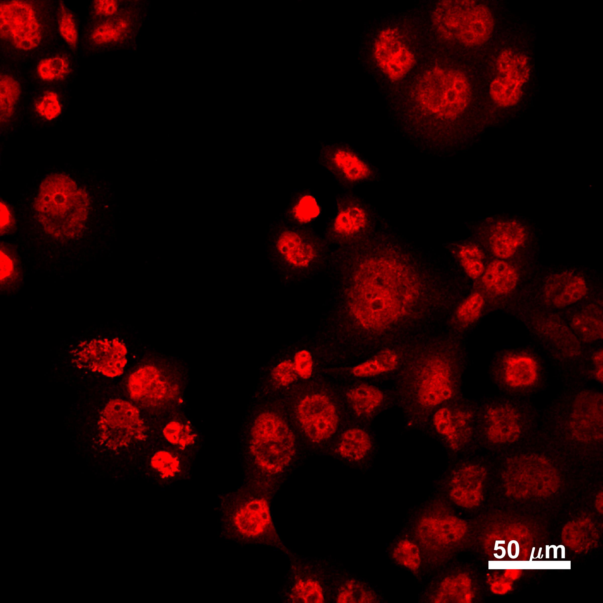
 fixed HepG2 cells using TIA1 antibody (12133-2-AP) at dilution of 1:200 and CoraLite®488-Conjugated Goat Anti-Rabbit IgG(H+L).")
 fixed L02 cells using TIA1 antibody (12133-2-AP) at dilution of 1:200 and CoraLite®488-Conjugated Goat Anti-Rabbit IgG(H+L).")
 fixed HeLa cells using 12133-2-AP (TIA1 antibody) at dilution of 1:50 and CoraLite488-Conjugated Goat Anti-Rabbit IgG(H+L).")
 and CoraLite®488-Conjugated Goat Anti-Rabbit IgG(H+L) at dilution 1:1000 (red), or 0.4 ug Isotype Control. Cells were fixed and permeabilized with Transcription Factor Staining Buffer Kit (PF00011).")
Applications testées
| Résultats positifs en WB | cellules COLO 320, cellules Jurkat, cellules Raji, tissu de thymus de souris, tissu splénique de souris |
| Résultats positifs en IP | cellules Jurkat |
| Résultats positifs en IHC | tissu de lymphome humain, tissu cérébral de souris il est suggéré de démasquer l'antigène avec un tampon de TE buffer pH 9.0; (*) À défaut, 'le démasquage de l'antigène peut être 'effectué avec un tampon citrate pH 6,0. |
| Résultats positifs en IF/ICC | cellules L02, cellules HeLa, cellules HepG2 |
| Résultats positifs en FC (Intra) | cellules HeLa, |
Dilution recommandée
| Application | Dilution |
|---|---|
| Western Blot (WB) | WB : 1:500-1:3000 |
| Immunoprécipitation (IP) | IP : 0.5-4.0 ug for 1.0-3.0 mg of total protein lysate |
| Immunohistochimie (IHC) | IHC : 1:50-1:500 |
| Immunofluorescence (IF)/ICC | IF/ICC : 1:50-1:500 |
| Flow Cytometry (FC) (INTRA) | FC (INTRA) : 0.40 ug per 10^6 cells in a 100 µl suspension |
| It is recommended that this reagent should be titrated in each testing system to obtain optimal results. | |
| Sample-dependent, check data in validation data gallery | |
Applications publiées
| KD/KO | See 6 publications below |
| WB | See 41 publications below |
| IHC | See 4 publications below |
| IF | See 39 publications below |
| CoIP | See 1 publications below |
| ChIP | See 1 publications below |
| RIP | See 1 publications below |
Informations sur le produit
12133-2-AP cible TIA1 dans les applications de WB, IHC, IF/ICC, FC (Intra), IP, CoIP, ChIP, RIP, ELISA et montre une réactivité avec des échantillons Humain, rat, souris
| Réactivité | Humain, rat, souris |
| Réactivité citée | rat, Humain, porc, poulet, souris |
| Hôte / Isotype | Lapin / IgG |
| Clonalité | Polyclonal |
| Type | Anticorps |
| Immunogène | TIA1 Protéine recombinante Ag2778 |
| Nom complet | TIA1 cytotoxic granule-associated RNA binding protein |
| Masse moléculaire calculée | 214 aa, 24 kDa, 43 kDa |
| Poids moléculaire observé | ~40 kDa |
| Numéro d’acquisition GenBank | BC015944 |
| Symbole du gène | TIA1 |
| Identification du gène (NCBI) | 7072 |
| Conjugaison | Non conjugué |
| Forme | Liquide |
| Méthode de purification | Purification par affinité contre l'antigène |
| Tampon de stockage | PBS with 0.02% sodium azide and 50% glycerol |
| Conditions de stockage | Stocker à -20°C. Stable pendant un an après l'expédition. L'aliquotage n'est pas nécessaire pour le stockage à -20oC Les 20ul contiennent 0,1% de BSA. |
Informations générales
TIA1, also named as p40-TIA-1, is involved in alternative pre-RNA splicing and regulation of mRNA translation by binding to AU-rich elements (AREs) located in mRNA 3' untranslated regions (3' UTRs). It possesses nucleolytic activity against cytotoxic lymphocyte target cells. TIA1 may be involved in apoptosis. Two isoforms of this protein exist - 41kDa and 42kDa. one of these was a missense variant (P362L) in TIA1. Similar to the ALS-related disease proteins TDP-43, hnRNPA1, and FUS, TIA1 is an RNA-binding protein containing a prionlike LCD and assembles into membrane-less organelles, including SGs. Postmortem neuropathology of five TIA1mutations carriers showed a consistent pathological signature with numerous round, hyaline, TAR DNA-binding protein 43 (TDP-43)-positive inclusions.TIA1mutations significantly increased the propensity of TIA1 protein to undergo phase transition. In live cells,TIA1mutations delayed stress granule (SG) disassembly and promoted the accumulation of non-dynamic SGs that harbored TDP-43. Moreover, TDP-43 in SGs became less mobile and insoluble.
Protocole
| Product Specific Protocols | |
|---|---|
| WB protocol for TIA1 antibody 12133-2-AP | Download protocol |
| IHC protocol for TIA1 antibody 12133-2-AP | Download protocol |
| IF protocol for TIA1 antibody 12133-2-AP | Download protocol |
| IP protocol for TIA1 antibody 12133-2-AP | Download protocol |
| Standard Protocols | |
|---|---|
| Click here to view our Standard Protocols |
Publications
| Species | Application | Title |
|---|---|---|
Cell In vivo structural characterization of the SARS-CoV-2 RNA genome identifies host proteins vulnerable to repurposed drugs.
| ||
Cell Res Predicting dynamic cellular protein-RNA interactions by deep learning using in vivo RNA structures. | ||
Nat Struct Mol Biol TDP-43 aggregation induced by oxidative stress causes global mitochondrial imbalance in ALS. | ||
Neuron TIA1 Mutations in Amyotrophic Lateral Sclerosis and Frontotemporal Dementia Promote Phase Separation and Alter Stress Granule Dynamics. | ||
Nat Commun Cellular stress alters 3'UTR landscape through alternative polyadenylation and isoform-specific degradation. | ||
Brain Defective cyclophilin A induces TDP-43 proteinopathy: implications for amyotrophic lateral sclerosis and frontotemporal dementia |
Avis
The reviews below have been submitted by verified Proteintech customers who received an incentive for providing their feedback.
FH haibo (Verified Customer) (09-28-2020) | Western blotting was good. But can not see clear granule signal even after treatment by IF. The cells used were fibroblasts.
|
FH Manohar (Verified Customer) (09-23-2020) | Used 1% Milk for blocking
|
FH Joshua (Verified Customer) (12-28-2019) | PANC1 cells fixed in 4% paraformaldehyde and stained overnight at 4C. Bright staining, mix of nuclear and stress granule localization as expected.
 |
FH Joshua (Verified Customer) (12-12-2018) | We found acetone fixation (20 min in 100% acetone at -20C) worked better than paraformaldehyde.
 |

