- Phare
- Validé par KD/KO
Anticorps Polyclonal de lapin anti-TNFR1/CD120a
TNFR1/CD120a Polyclonal Antibody for WB, IHC, IF-P, ELISA
Hôte / Isotype
Lapin / IgG
Réactivité testée
Humain, souris et plus (2)
Applications
WB, IHC, IF-P, IP, CoIP, ELISA
Conjugaison
Non conjugué
N° de cat : 21574-1-AP
Synonymes
Galerie de données de validation
 at dilution of 1:600 incubated at room temperature for 1.5 hours.")
 at dilution of 1:500 incubated at room temperature for 1.5 hours.")
 at dilution of 1:500 incubated at room temperature for 1.5 hours.")
 at dilution of 1:600 incubated at room temperature for 1.5 hours.")
 at dilution of 1:200 (under 10x lens. Heat mediated antigen retrieval with Tris-EDTA buffer (pH 9.0).")
 at dilution of 1:200 (under 40x lens. Heat mediated antigen retrieval with Tris-EDTA buffer (pH 9.0).")
 at dilution of 1:200 (under 10x lens). Heat mediated antigen retrieval with Tris-EDTA buffer (pH 9.0).")
 at dilution of 1:200 (under 40x lens). Heat mediated antigen retrieval with Tris-EDTA buffer (pH 9.0).")
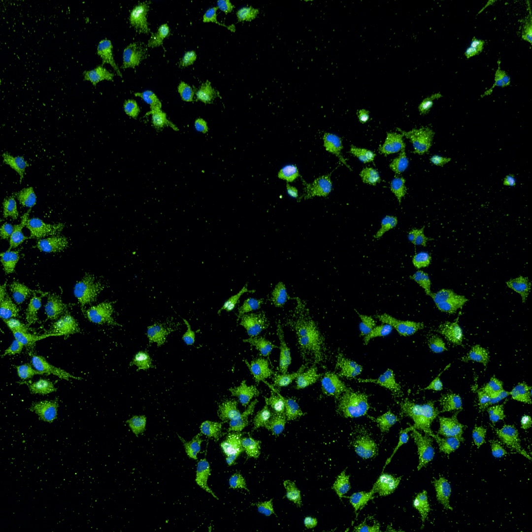
 fixed mouse brain tissue using TNFR1/CD120a antibody (21574-1-AP) at dilution of 1:200 and CoraLite®488-Conjugated AffiniPure Goat Anti-Rabbit IgG(H+L).")
 fixed mouse brain tissue using TNFR1 antibody (21574-1-AP) at dilution of 1:200 and CoraLite®488-Conjugated AffiniPure Goat Anti-Rabbit IgG(H+L).")
Applications testées
| Résultats positifs en WB | cellules Raji, cellules HeLa, cellules HL-60, tissu cérébral humain |
| Résultats positifs en IHC | tissu cérébral humain, tissu de cancer du sein humain il est suggéré de démasquer l'antigène avec un tampon de TE buffer pH 9.0; (*) À défaut, 'le démasquage de l'antigène peut être 'effectué avec un tampon citrate pH 6,0. |
| Résultats positifs en IF-P | tissu cérébral de souris, |
Dilution recommandée
| Application | Dilution |
|---|---|
| Western Blot (WB) | WB : 1:500-1:1000 |
| Immunohistochimie (IHC) | IHC : 1:50-1:500 |
| Immunofluorescence (IF)-P | IF-P : 1:50-1:500 |
| It is recommended that this reagent should be titrated in each testing system to obtain optimal results. | |
| Sample-dependent, check data in validation data gallery | |
Applications publiées
| KD/KO | See 3 publications below |
| WB | See 62 publications below |
| IHC | See 11 publications below |
| IF | See 17 publications below |
| IP | See 3 publications below |
| CoIP | See 1 publications below |
Informations sur le produit
21574-1-AP cible TNFR1/CD120a dans les applications de WB, IHC, IF-P, IP, CoIP, ELISA et montre une réactivité avec des échantillons Humain, souris
| Réactivité | Humain, souris |
| Réactivité citée | rat, Humain, porc, souris |
| Hôte / Isotype | Lapin / IgG |
| Clonalité | Polyclonal |
| Type | Anticorps |
| Immunogène | TNFR1/CD120a Protéine recombinante Ag16112 |
| Nom complet | tumor necrosis factor receptor superfamily, member 1A |
| Masse moléculaire calculée | 455 aa, 50 kDa |
| Poids moléculaire observé | 50 kDa |
| Numéro d’acquisition GenBank | BC010140 |
| Symbole du gène | TNFR1 |
| Identification du gène (NCBI) | 7132 |
| Conjugaison | Non conjugué |
| Forme | Liquide |
| Méthode de purification | Purification par affinité contre l'antigène |
| Tampon de stockage | PBS with 0.02% sodium azide and 50% glycerol |
| Conditions de stockage | Stocker à -20°C. Stable pendant un an après l'expédition. L'aliquotage n'est pas nécessaire pour le stockage à -20oC Les 20ul contiennent 0,1% de BSA. |
Informations générales
Tumor necrosis factor (TNF) is a multifunctional cytokine that plays a key role in regulating inflammation, immune functions, host defense, and apoptosis (PMID: 16407280). TNF exists in soluble and membrane-bound forms. TNF signals through two distinct cell surface receptors, TNFR1 (TNFRSF1A, CD120a) and TNFR2 (TNFRSF1B, CD120b). Whereas TNFR1 is widely expressed, expression of TNFR2 is limited to cells of the immune system, endothelial cells, and nerve cells (PMID: 22053109). TNFR1, which contains a death domain (DD) within its intracytoplasmic region, is thought to be the key receptor for TNF signaling (PMID: 16407280). This receptor can activate NF-kappaB, mediate apoptosis, and function as a regulator of inflammation. Antiapoptotic protein BCL2-associated athanogene 4 (BAG4/SODD) and adaptor proteins TRADD and TRAF2 have been shown to interact with this receptor, and thus play regulatory roles in the signal transduction mediated by the receptor.
Protocole
| Product Specific Protocols | |
|---|---|
| WB protocol for TNFR1/CD120a antibody 21574-1-AP | Download protocol |
| IHC protocol for TNFR1/CD120a antibody 21574-1-AP | Download protocol |
| IF protocol for TNFR1/CD120a antibody 21574-1-AP | Download protocol |
| Standard Protocols | |
|---|---|
| Click here to view our Standard Protocols |
Publications
| Species | Application | Title |
|---|---|---|
Cancer Commun (Lond) Targeting autophagy overcomes cancer-intrinsic resistance to CAR-T immunotherapy in B-cell malignancies | ||
Adv Healthc Mater Hyaluronic Acid-based ROS-responsive Multifunctional Injectable Hydrogel Platform Accelerating Diabetic Wound Healing | ||
Aging Dis MiR-29a-3p Improves Acute Lung Injury by Reducing Alveolar Epithelial Cell PANoptosis. | ||
PLoS Biol A unique death pathway keeps RIPK1 D325A mutant mice in check at embryonic day 10.5. | ||
Cancer Lett Up-regulation of OLR1 expression by TBC1D3 through activation of TNFα/NF-κB pathway promotes the migration of human breast cancer cells. |
Avis
The reviews below have been submitted by verified Proteintech customers who received an incentive for providing their feedback.
FH Xinda (Verified Customer) (08-16-2024) | It gave very nice and specific bands.
|
FH Tinne (Verified Customer) (08-14-2023) | This AB worked quite well - not the strongest signal - might be worth increasing the concentration. I got the best results when I did not permeabilise the cells.
 |

