- Phare
- Validé par KD/KO
Anticorps Polyclonal de lapin anti-TNFR2/CD120b
TNFR2/CD120b Polyclonal Antibody for WB, IHC, IF/ICC, IP, ELISA
Hôte / Isotype
Lapin / IgG
Réactivité testée
Humain, rat, souris
Applications
WB, IHC, IF/ICC, IP, CoIP, ELISA
Conjugaison
Non conjugué
N° de cat : 19272-1-AP
Synonymes
Galerie de données de validation
 at dilution of 1:500 incubated at room temperature for 1.5 hours.")
 at dilution of 1:600 incubated at room temperature for 1.5 hours.")
 at dilution of 1:500 incubated at room temperature for 1.5 hours.")
 with HEK-293 cells lysate 1200ug.")
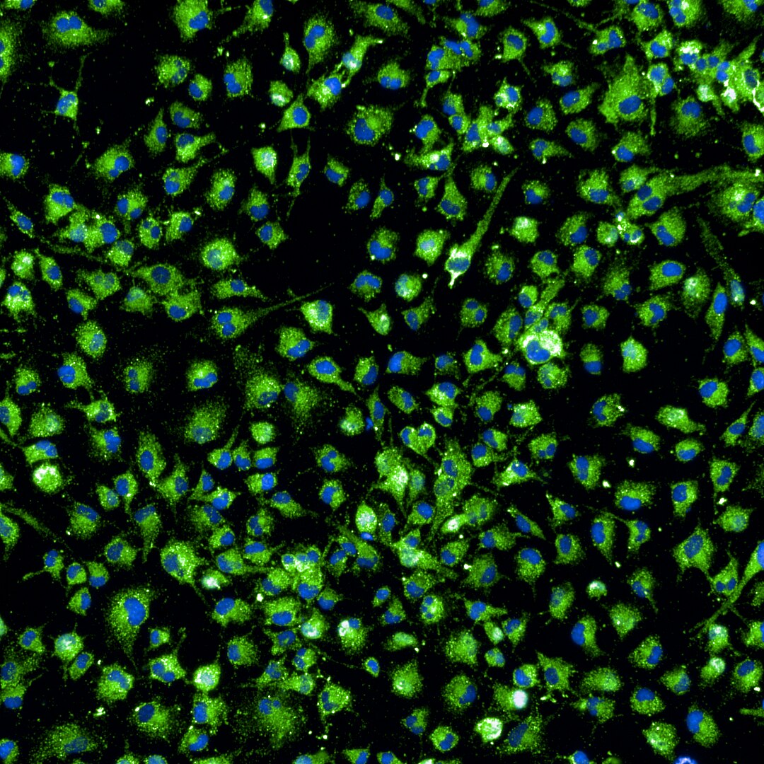
 at dilution of 1:200 (under 10x lens).")
 at dilution of 1:200 (under 40x lens).")
 fixed THP-1 cells using TNFR2 / TNFRSF1B antibody (19272-1-AP) at dilution of 1:200 and CoraLite®488-Conjugated AffiniPure Goat Anti-Rabbit IgG(H+L) (SA00013-2).")
Applications testées
| Résultats positifs en WB | NK-92 cells, cellules HEK-293, cellules Jurkat, cellules THP-1, tissu de thymus de rat, tissu de thymus de souris |
| Résultats positifs en IP | cellules HEK-293 |
| Résultats positifs en IHC | tissu splénique humain, il est suggéré de démasquer l'antigène avec un tampon de TE buffer pH 9.0; (*) À défaut, 'le démasquage de l'antigène peut être 'effectué avec un tampon citrate pH 6,0. |
| Résultats positifs en IF/ICC | cellules THP-1, |
Dilution recommandée
| Application | Dilution |
|---|---|
| Western Blot (WB) | WB : 1:500-1:1000 |
| Immunoprécipitation (IP) | IP : 0.5-4.0 ug for 1.0-3.0 mg of total protein lysate |
| Immunohistochimie (IHC) | IHC : 1:50-1:500 |
| Immunofluorescence (IF)/ICC | IF/ICC : 1:50-1:500 |
| It is recommended that this reagent should be titrated in each testing system to obtain optimal results. | |
| Sample-dependent, check data in validation data gallery | |
Applications publiées
| KD/KO | See 4 publications below |
| WB | See 30 publications below |
| IHC | See 9 publications below |
| IF | See 13 publications below |
| IP | See 1 publications below |
| CoIP | See 1 publications below |
Informations sur le produit
19272-1-AP cible TNFR2/CD120b dans les applications de WB, IHC, IF/ICC, IP, CoIP, ELISA et montre une réactivité avec des échantillons Humain, rat, souris
| Réactivité | Humain, rat, souris |
| Réactivité citée | rat, Humain, souris |
| Hôte / Isotype | Lapin / IgG |
| Clonalité | Polyclonal |
| Type | Anticorps |
| Immunogène | TNFR2/CD120b Protéine recombinante Ag5866 |
| Nom complet | tumor necrosis factor receptor superfamily, member 1B |
| Masse moléculaire calculée | 48 kDa |
| Poids moléculaire observé | 70-75 kDa |
| Numéro d’acquisition GenBank | BC052977 |
| Symbole du gène | TNFR2 |
| Identification du gène (NCBI) | 7133 |
| Conjugaison | Non conjugué |
| Forme | Liquide |
| Méthode de purification | Purification par affinité contre l'antigène |
| Tampon de stockage | PBS with 0.02% sodium azide and 50% glycerol |
| Conditions de stockage | Stocker à -20°C. Stable pendant un an après l'expédition. L'aliquotage n'est pas nécessaire pour le stockage à -20oC Les 20ul contiennent 0,1% de BSA. |
Informations générales
Tumor necrosis factor-alpha (TNFA/TNFSF2) is a multifunctional cytokine that plays a key role in regulating inflammation, immune functions, host defense, and apoptosis (PMID: 16407280). TNFA signals through two distinct cell surface receptors, TNFR1 (TNFRSF1A, CD120a, p55) and TNFR2 (TNFRSF1B, CD120b, p75). TNFR1 is widely expressed, whereas TNFR2 exhibits more restricted expression, being found on CD4 and CD8 T lymphocytes, endothelial cells, microglia, oligodendrocytes, neuron subtypes, cardiac myocytes, thymocytes and human mesenchymal stem cells (PMID: 20489699; 22374304). In contrast to TNFR1, TNFR2 does not have a death domain. TNFR2 only signals for antiapoptotic reactions. However, recent evidence indicates that TNFR2 also signals to induce TRAF2 degradation (PMID: 22374304). Various defects in the TNFR2 pathway, due to polymorphisms in the TNFR2 gene, upregulated expression of TNFR2 and TNFR2 shedding, have been implicated in the pathology of several autoimmune disorders (PMID: 20489699).
Protocole
| Product Specific Protocols | |
|---|---|
| WB protocol for TNFR2/CD120b antibody 19272-1-AP | Download protocol |
| IHC protocol for TNFR2/CD120b antibody 19272-1-AP | Download protocol |
| IF protocol for TNFR2/CD120b antibody 19272-1-AP | Download protocol |
| IP protocol for TNFR2/CD120b antibody 19272-1-AP | Download protocol |
| Standard Protocols | |
|---|---|
| Click here to view our Standard Protocols |
Publications
| Species | Application | Title |
|---|---|---|
Nat Immunol NKILA lncRNA promotes tumor immune evasion by sensitizing T cells to activation-induced cell death. | ||
Mol Ther Tumor necrosis factor alpha delivers exogenous inflammation-related microRNAs to recipient cells with functional targeting capabilities. | ||
Mater Today Bio Intracellular hydrogelation of macrophage conjugated probiotics for hitchhiking delivery and combined treatment of colitis | ||
Oxid Med Cell Longev Protective Effects of Cinnamaldehyde against Mesenteric Ischemia-Reperfusion-Induced Lung and Liver Injuries in Rats. | ||
Cancer Lett Up-regulation of OLR1 expression by TBC1D3 through activation of TNFα/NF-κB pathway promotes the migration of human breast cancer cells. | ||
Pain Nox2-dependent signaling between macrophages and sensory neurons contributes to neuropathic pain hypersensitivity. |
Avis
The reviews below have been submitted by verified Proteintech customers who received an incentive for providing their feedback.
FH Tinne (Verified Customer) (08-14-2023) | This worked really well for ICC of human HPCs.
 |

