- Phare
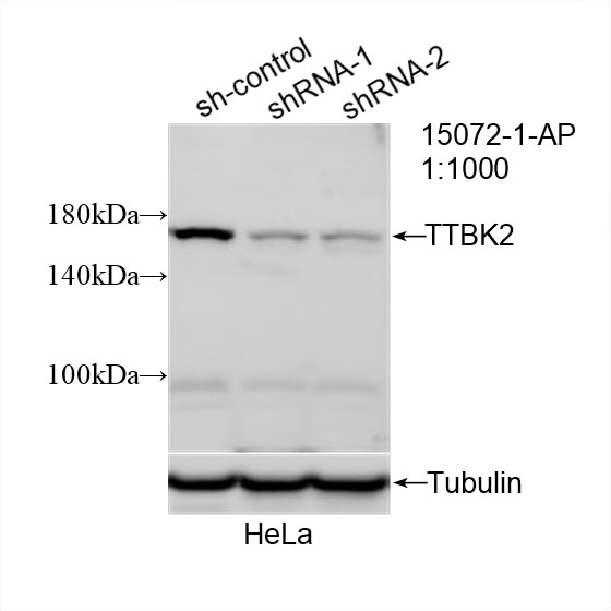
- Validé par KD/KO
Anticorps Polyclonal de lapin anti-TTBK2
TTBK2 Polyclonal Antibody for WB, IHC, IF/ICC, ELISA
Hôte / Isotype
Lapin / IgG
Réactivité testée
Humain, rat, souris
Applications
WB, IHC, IF/ICC, ELISA
Conjugaison
Non conjugué
N° de cat : 15072-1-AP
Synonymes
Galerie de données de validation
Applications testées
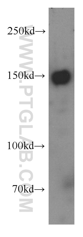
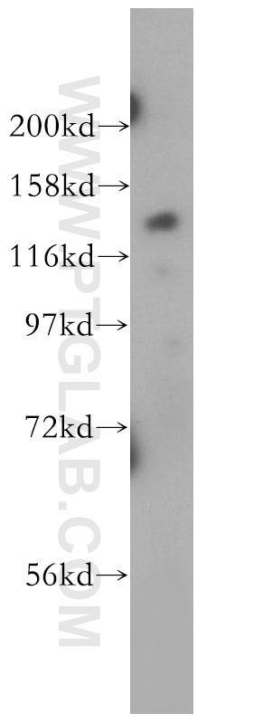
| Résultats positifs en WB | cellules PC-3, cellules HeLa, cellules SH-SY5Y |
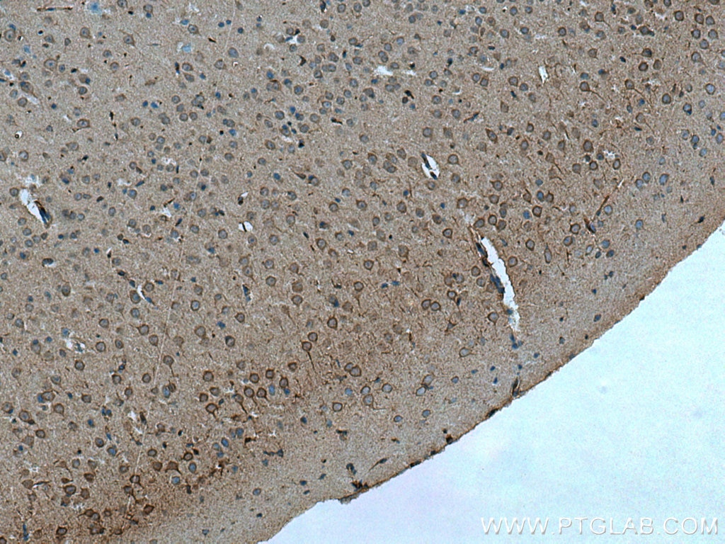
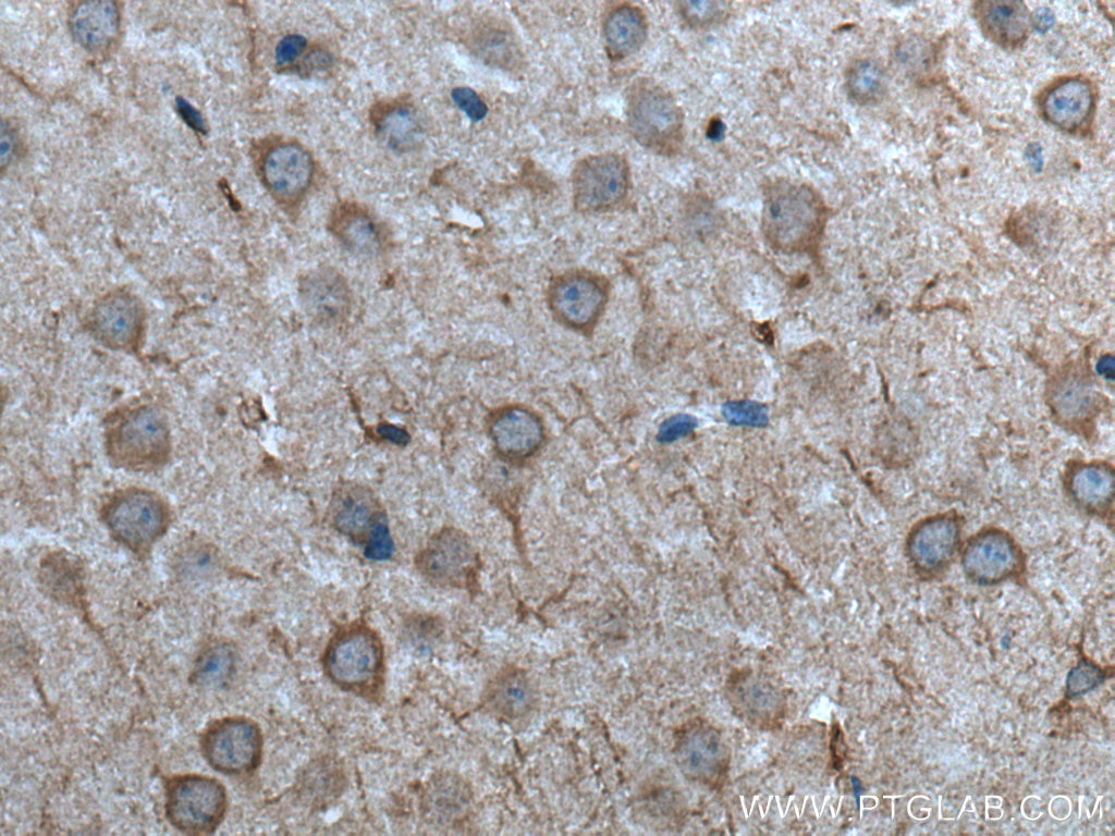
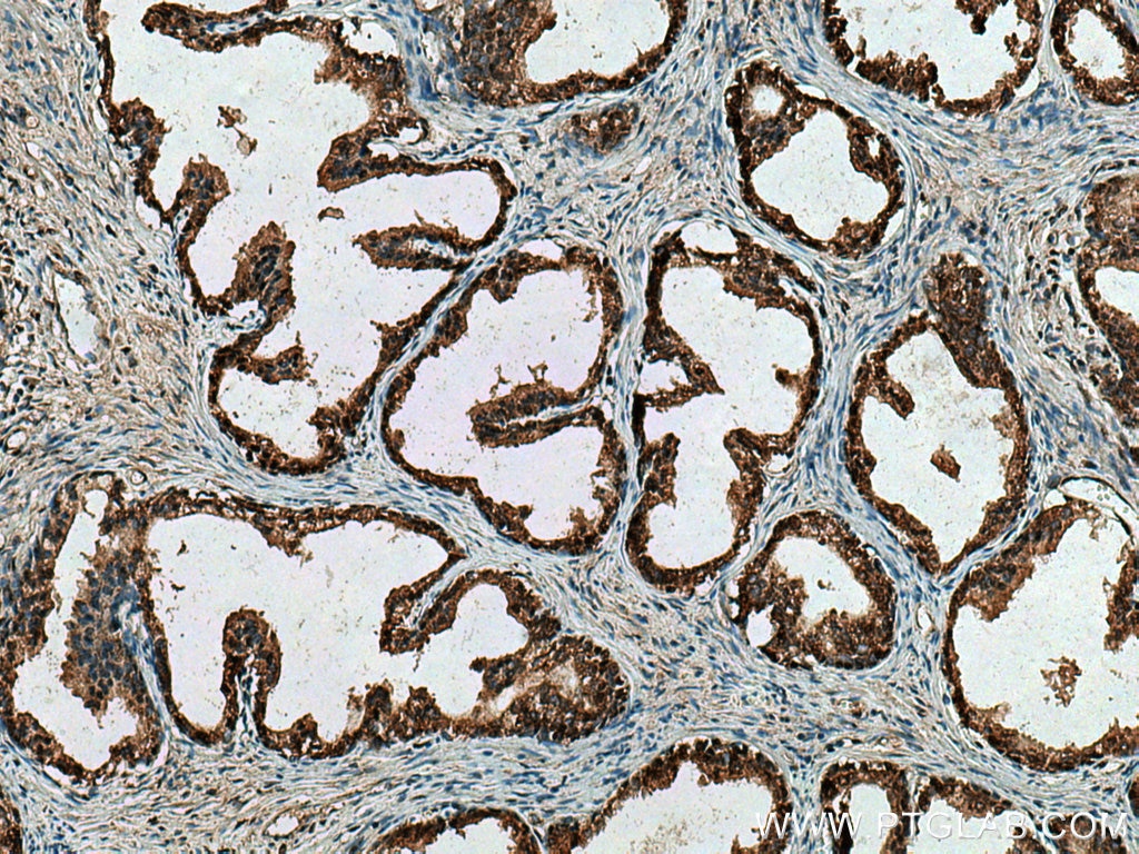
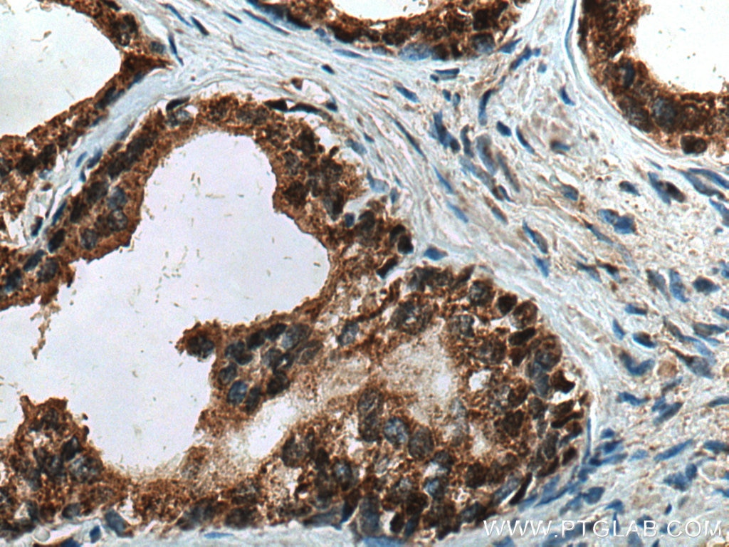
| Résultats positifs en IHC | tissu cérébral de souris, tissu de cancer de la prostate humain il est suggéré de démasquer l'antigène avec un tampon de TE buffer pH 9.0; (*) À défaut, 'le démasquage de l'antigène peut être 'effectué avec un tampon citrate pH 6,0. |
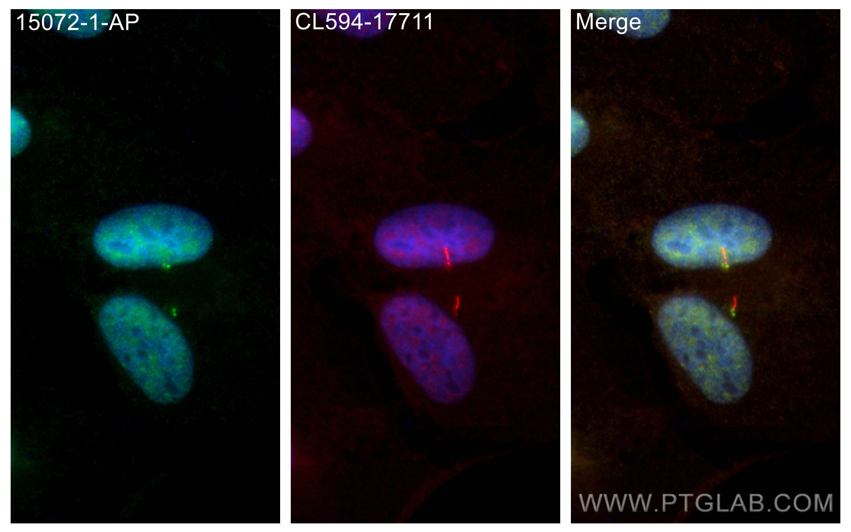
| Résultats positifs en IF/ICC | cellules hTERT-RPE1, |
Dilution recommandée
| Application | Dilution |
|---|---|
| Western Blot (WB) | WB : 1:500-1:2000 |
| Immunohistochimie (IHC) | IHC : 1:50-1:500 |
| Immunofluorescence (IF)/ICC | IF/ICC : 1:200-1:800 |
| It is recommended that this reagent should be titrated in each testing system to obtain optimal results. | |
| Sample-dependent, check data in validation data gallery | |
Applications publiées
| WB | See 3 publications below |
| IF | See 6 publications below |
Informations sur le produit
15072-1-AP cible TTBK2 dans les applications de WB, IHC, IF/ICC, ELISA et montre une réactivité avec des échantillons Humain, rat, souris
| Réactivité | Humain, rat, souris |
| Réactivité citée | Humain, souris |
| Hôte / Isotype | Lapin / IgG |
| Clonalité | Polyclonal |
| Type | Anticorps |
| Immunogène | TTBK2 Protéine recombinante Ag7108 |
| Nom complet | tau tubulin kinase 2 |
| Masse moléculaire calculée | 137 kDa |
| Poids moléculaire observé | 137-150 kDa |
| Numéro d’acquisition GenBank | BC071556 |
| Symbole du gène | TTBK2 |
| Identification du gène (NCBI) | 146057 |
| Conjugaison | Non conjugué |
| Forme | Liquide |
| Méthode de purification | Purification par affinité contre l'antigène |
| Tampon de stockage | PBS with 0.02% sodium azide and 50% glycerol |
| Conditions de stockage | Stocker à -20°C. Stable pendant un an après l'expédition. L'aliquotage n'est pas nécessaire pour le stockage à -20oC Les 20ul contiennent 0,1% de BSA. |
Informations générales
TTBK2(tau tubulin kinase 2) plays an important role in the tau cascade and in spinocerebellar degeneration. The N-terminus of TTBK2 has a serine-threonine-tyrosine kinase domain, and a C-terminal region shows homology to TTBK1. TTBK1 and TTBK2 function as regulators of TDP-43 phosphorylation. TTBK1/2 may be attractive drug targets for therapeutic interventions in TDP-43 proteinopathies such as FTLD-TDP and ALS.(PMID:25473830). 15072-1-AP has a cross reaction with TTBK1.
Protocole
| Product Specific Protocols | |
|---|---|
| WB protocol for TTBK2 antibody 15072-1-AP | Download protocol |
| IHC protocol for TTBK2 antibody 15072-1-AP | Download protocol |
| IF protocol for TTBK2 antibody 15072-1-AP | Download protocol |
| Standard Protocols | |
|---|---|
| Click here to view our Standard Protocols |
Publications
| Species | Application | Title |
|---|---|---|
Cell Res NudCL2 is an autophagy receptor that mediates selective autophagic degradation of CP110 at mother centrioles to promote ciliogenesis. | ||
EMBO Rep XIAP-mediated degradation of IFT88 disrupts HSC cilia to stimulate HSC activation and liver fibrosis | ||
Sci Rep MCRS1 associates with cytoplasmic dynein and mediates pericentrosomal material recruitment. | ||
Hum Mol Genet Altered gene regulation as a candidate mechanism by which ciliopathy gene SDCCAG8 contributes to schizophrenia and cognitive function. | ||
J Cell Sci Differential requirements for the EF-hands of human centrin2 in primary ciliogenesis and nucleotide excision repair. |
Avis
The reviews below have been submitted by verified Proteintech customers who received an incentive for providing their feedback.
FH Christine (Verified Customer) (01-27-2023) | Used on lysates from HEK293T cells. Incubation for a few hours at room temperature. Detection of 2 main bands, 1 above 118 kDa marker (expected size of TTBK2 is 137-150 kDa) and 1 between 47 and 85 kDa markers.
|
