- Phare
- Validé par KD/KO
Anticorps Monoclonal anti-TUBB3-specific/TUJ1
TUBB3-specific/TUJ1 Monoclonal Antibody for WB, IHC, IF/ICC, IF-P, IF-Fro, FC (Intra), IP, ELISA
Hôte / Isotype
Mouse / IgG1
Réactivité testée
Humain, Lapin, porc, poulet, rat, souris
Applications
WB, IHC, IF/ICC, IF-P, IF-Fro, FC (Intra), IP, ELISA
Conjugaison
Non conjugué
CloneNo.
1F8G10
N° de cat : 66375-1-Ig
Synonymes
Galerie de données de validation
 at dilution of 1:49000 incubated at room temperature for 1.5 hours.")
 with sh-Control and sh-TUBB3-specific/TUJ1 transfected HEK-293 cells.")
 at dilution of 1:40,000 incubated at room temperature for 1.5 hours.")
 with SH-SY5Y cells lysate 1240 ug.")
 at dilution of 1:200 (under 40x lens. Heat mediated antigen retrieval with Tris-EDTA buffer (pH 9.0).")
 at dilution of 1:400 (under 10x lens).")
 at dilution of 1:400 (under 40x lens).")
 at dilution of 1:20000 (under 10x lens). Heat mediated antigen retrieval with Tris-EDTA buffer (pH 9.0).")
 at dilution of 1:20000 (under 40x lens). Heat mediated antigen retrieval with Tris-EDTA buffer (pH 9.0).")
 at dilution of 1:400 (under 10x lens. Heat mediated antigen retrieval with Tris-EDTA buffer (pH 9.0).")
 at dilution of 1:400 (under 40x lens. Heat mediated antigen retrieval with Tris-EDTA buffer (pH 9.0).")
 at dilution of 1:200 (under 10x lens. Heat mediated antigen retrieval with Tris-EDTA buffer (pH 9.0).")
 at dilution of 1:200 (under 0x lens. Heat mediated antigen retrieval with Tris-EDTA buffer (pH 9.0).")
 generated from human induced pluripotent stem cells (iPSCs) and fixed with 4% PFA. Stained for Tubulin beta 3/TUJ1 using 66375-1-Ig at 1:500 dilution (green) and Cytokeratin 19 using 10712-1-AP at 1:200 (red). Nuclear stain DAPI (blue). Scale bar = 100 µm. Data generated by Alessandro Bellapianta at Johannes Kepler Universitat, Austria.")
 generated from human induced pluripotent stem cells (iPSCs) and fixed with 4% PFA. Stained for Tubulin beta 3/TUJ1 using 66375-1-Ig at 1:500 dilution (green) and CRALBP using 15356-1-AP at 1:400 (red). Nuclear stain DAPI (blue). Scale bar = 50 µm. Data generated by Alessandro Bellapianta at Johannes Kepler Universitat, Austria.")
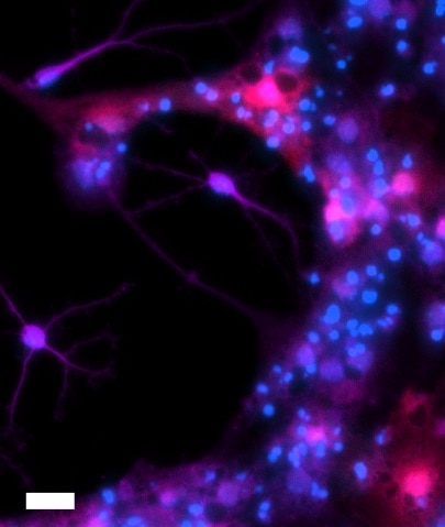
 at 1/200 (Magenta) and neurons with TUJ1 (66375-1-Ig) at 1:500 (Green). The sample was fixed with 4% Paraformaldehyde and permeabilized with 0.3% Triton X-100. Alexa Fluor 488-conjugated goat anti-mouse IgG (1/500) and Alexa Fluor 594-conjugated goat anti-rabbit IgG (1/500) were used as the secondary antibodies. Nuclei were counterstained with DAPI (blue).")
 generated from human induced pluripotent stem cells (iPSCs) and fixed with 4% PFA. Stained for Tubulin beta 3/TUJ1 using 66375-1-Ig at 1:500 dilution (green) and PAX6 (12323-1-AP) at 1:500. Nuclear stain DAPI (blue). Scale bar = 50 µm. Data generated by Alessandro Bellapianta at Johannes Kepler Universitat, Austria.")
 fixed rat brain tissue using 66375-1-Ig (TUBB3-specific antibody), at dilution of 1:200 and CoraLite®488-Conjugated AffiniPure Goat Anti-Mouse IgG(H+L). The section was co-stained with 26975-1-AP (NeuN antibody, red).")
 fixed mouse brain tissue using 66375-1-Ig (TUBB3-specific antibody) at dilution of 1:50 and Alexa Fluor 488-Conjugated AffiniPure Goat Anti-Mouse IgG(H+L).")
 fixed mouse brain tissue using 66375-1-Ig (TUBB3-specific antibody) at dilution of 1:50 and Alexa Fluor 488-Conjugated AffiniPure Goat Anti-Mouse IgG(H+L).")
 fixed frozen OCT-embedded rat brain tissue using TUBB3-specific/TUJ1 antibody (66375-1-Ig, Clone: 1F8G10 ) at dilution of 1:800 and CoraLite®488-Conjugated Goat Anti-Mouse IgG(H+L) (SA00013-1).")
 fixed frozen OCT-embedded rat cerebellum tissue using TUBB3-specific/TUJ1 antibody (66375-1-Ig, Clone: 1F8G10 ) at dilution of 1:400 and CoraLite®488-Conjugated Goat Anti-Mouse IgG(H+L) (SA00013-1).")
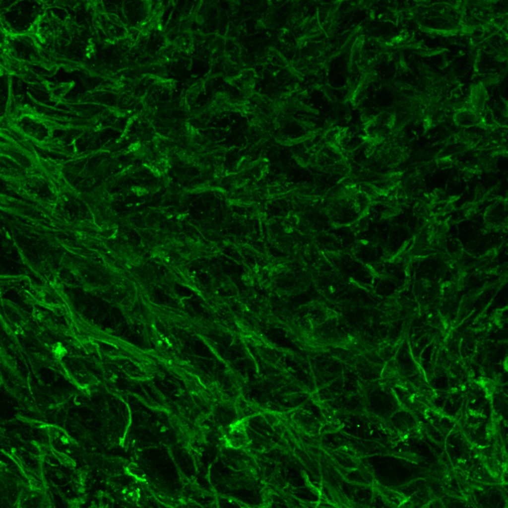
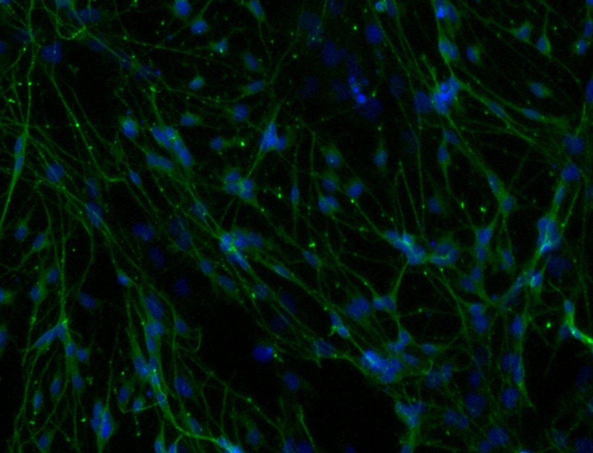
 with 4% PFA fixed control hiPSC derived neuronal cultures (35 days old). (Green: TUBB3; Blue: DAPI). Provided by BioTalentum Ltd., Hungary.")
 and CoraLite®488-Conjugated AffiniPure Goat Anti-Mouse IgG(H+L) at dilution 1:1000 (red), or 0.2 ug Control Antibody. Cells were fixed with 4% PFA and permeabilized with Flow Cytometry Perm Buffer (PF00011-C).")
Applications testées
| Résultats positifs en WB | cellules SH-SY5Y, cellules HEK-293, cellules Neuro-2a, cellules PC-12, cerveau de lapin, cerveau de porc, cerveau de poulet, tissu cérébral humain fœtal |
| Résultats positifs en IP | cellules SH-SY5Y, |
| Résultats positifs en IHC | tissu de cervelet humain, tissu cérébral de souris, tissu de cervelet de souris il est suggéré de démasquer l'antigène avec un tampon de TE buffer pH 9.0; (*) À défaut, 'le démasquage de l'antigène peut être 'effectué avec un tampon citrate pH 6,0. |
| Résultats positifs en IF-P | tissu cérébral de rat, tissu cérébral de souris |
| Résultats positifs en IF-Fro | tissu cérébral de rat, tissu de cervelet de rat |
| Résultats positifs en IF/ICC | cellules iPS, |
| Résultats positifs en FC (Intra) | cellules SH-SY5Y, |
Dilution recommandée
| Application | Dilution |
|---|---|
| Western Blot (WB) | WB : 1:5000-1:50000 |
| Immunoprécipitation (IP) | IP : 0.5-4.0 ug for 1.0-3.0 mg of total protein lysate |
| Immunohistochimie (IHC) | IHC : 1:400-1:20000 |
| Immunofluorescence (IF)-P | IF-P : 1:50-1:500 |
| Immunofluorescence (IF)-FRO | IF-FRO : 1:400-1:1600 |
| Immunofluorescence (IF)/ICC | IF/ICC : 1:125-1:500 |
| Flow Cytometry (FC) (INTRA) | FC (INTRA) : 0.20 ug per 10^6 cells in a 100 µl suspension |
| It is recommended that this reagent should be titrated in each testing system to obtain optimal results. | |
| Sample-dependent, check data in validation data gallery | |
Applications publiées
| WB | See 23 publications below |
| IHC | See 2 publications below |
| IF | See 62 publications below |
Informations sur le produit
66375-1-Ig cible TUBB3-specific/TUJ1 dans les applications de WB, IHC, IF/ICC, IF-P, IF-Fro, FC (Intra), IP, ELISA et montre une réactivité avec des échantillons Humain, Lapin, porc, poulet, rat, souris
| Réactivité | Humain, Lapin, porc, poulet, rat, souris |
| Réactivité citée | rat, Humain, poulet, souris |
| Hôte / Isotype | Mouse / IgG1 |
| Clonalité | Monoclonal |
| Type | Anticorps |
| Immunogène | Peptide |
| Nom complet | tubulin, beta 3 |
| Masse moléculaire calculée | 55 kDa |
| Poids moléculaire observé | 50-55 kDa |
| Numéro d’acquisition GenBank | NM_001197181 |
| Symbole du gène | TUBB3 |
| Identification du gène (NCBI) | 10381 |
| Conjugaison | Non conjugué |
| Forme | Liquide |
| Méthode de purification | Purification par protéine G |
| Tampon de stockage | PBS with 0.02% sodium azide and 50% glycerol |
| Conditions de stockage | Stocker à -20°C. Stable pendant un an après l'expédition. L'aliquotage n'est pas nécessaire pour le stockage à -20oC Les 20ul contiennent 0,1% de BSA. |
Informations générales
TUBB3, the class III β tubulin or Tuj1, is selectively expressed in testis and neurons of the central and peripheral nervous system. It has been widely used as a marker for neurons. Aberrant expression of TUBB3 has also been found in various tumors of non-neural origin and can be used as a biomarker for cancer aggressiveness and a marker for the tendency to respond poorly to chemotherapy. This antibody is specific to TUBB3 but not cross-react with other tubulin isoforms.
Protocole
| Product Specific Protocols | |
|---|---|
| WB protocol for TUBB3-specific/TUJ1 antibody 66375-1-Ig | Download protocol |
| IHC protocol for TUBB3-specific/TUJ1 antibody 66375-1-Ig | Download protocol |
| IF protocol for TUBB3-specific/TUJ1 antibody 66375-1-Ig | Download protocol |
| IP protocol for TUBB3-specific/TUJ1 antibody 66375-1-Ig | Download protocol |
| Standard Protocols | |
|---|---|
| Click here to view our Standard Protocols |
Publications
| Species | Application | Title |
|---|---|---|
Neuron Posterior Neocortex-Specific Regulation of Neuronal Migration by CEP85L Identifies Maternal Centriole-Dependent Activation of CDK5. | ||
Redox Biol LOX-mediated ECM mechanical stress induces Piezo1 activation in hypoxic-ischemic brain damage and identification of novel inhibitor of LOX | ||
Sci Total Environ Ambient NO2 exposure induces migraine in rats: Evidence, mechanisms and interventions. | ||
Curr Biol Pyramidal neuron morphogenesis requires a septin network that stabilizes filopodia and suppresses lamellipodia during neurite initiation | ||
Cell Rep NLRP14 deficiency causes female infertility with oocyte maturation defects and early embryonic arrest by impairing cytoplasmic UHRF1 abundance | ||
Theranostics Chlorogenic acid effectively treats cancers through induction of cancer cell differentiation. |
Avis
The reviews below have been submitted by verified Proteintech customers who received an incentive for providing their feedback.
FH Ankush (Verified Customer) (05-02-2025) | worked fine
 |
FH Gabriele (Verified Customer) (01-12-2023) | The antibody works very well with our IF protocol. Maybe the dilution could be pushed to 1:500-1:600. TUBB3 1:400 (green), Hoechst (Blue)
 |
FH Alessandro (Verified Customer) (07-27-2022) | No aspecific staining, great outcome
|
FH Josh (Verified Customer) (12-18-2018) | BIII tubulin antibody signal nicely fills neuronal cell bodies and processes with good signal to noise at a reasonable dilution (1:500 in 1% NDS, 0.3% Triton-X, PBS with donkey anti-mouse 647 secondary, 1:500). However, BIII tubulin antibody signal also colocalized with S100 immunoreactive astrocytic processes, though with a lesser intensity. Overall, the antibody is a useful marker of neuronal differentiation when used in combination with other cell-selective antibodies and morphology. Image caption: Differentiated adult hippocampal neural stem cell culture. BIII tubulin 1:500 (purple), S100 1:500 (red), Hoechst (blue). Scale bar = 20 um.
 |

