- Phare
- Validé par KD/KO
Anticorps Polyclonal de lapin anti-VAPA
VAPA Polyclonal Antibody for WB, IHC, IF/ICC, IP, ELISA
Hôte / Isotype
Lapin / IgG
Réactivité testée
Humain, rat, souris et plus (1)
Applications
WB, IHC, IF/ICC, IP, COIP, ELISA
Conjugaison
Non conjugué
N° de cat : 15275-1-AP
Synonymes
Galerie de données de validation
Applications testées
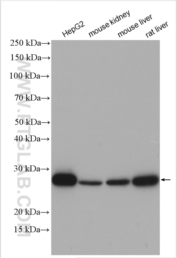
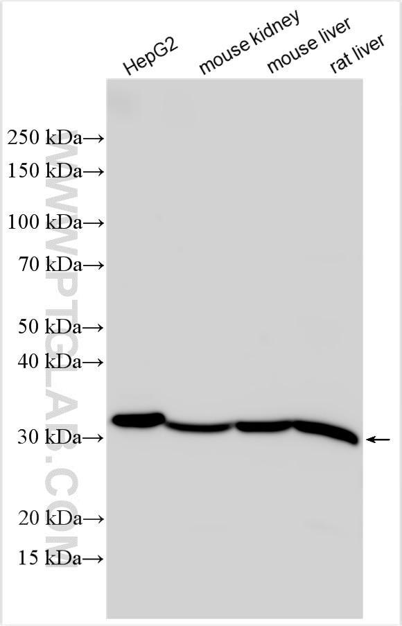
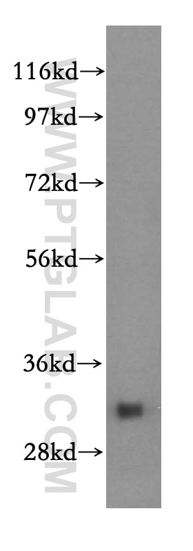
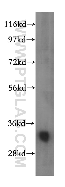
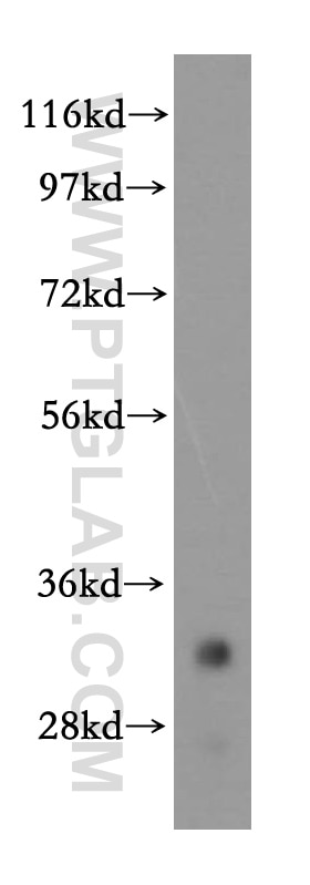
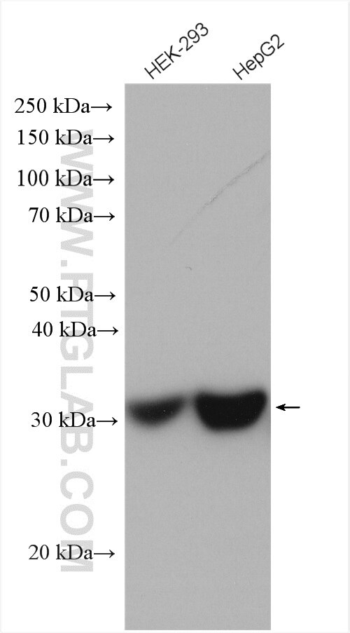
| Résultats positifs en WB | cellules HepG2, cellules HEK-293, tissu cérébral humain, tissu de muscle squelettique de souris, tissu hépatique de rat, tissu hépatique de souris, tissu rénal de souris, tissu rénal humain |
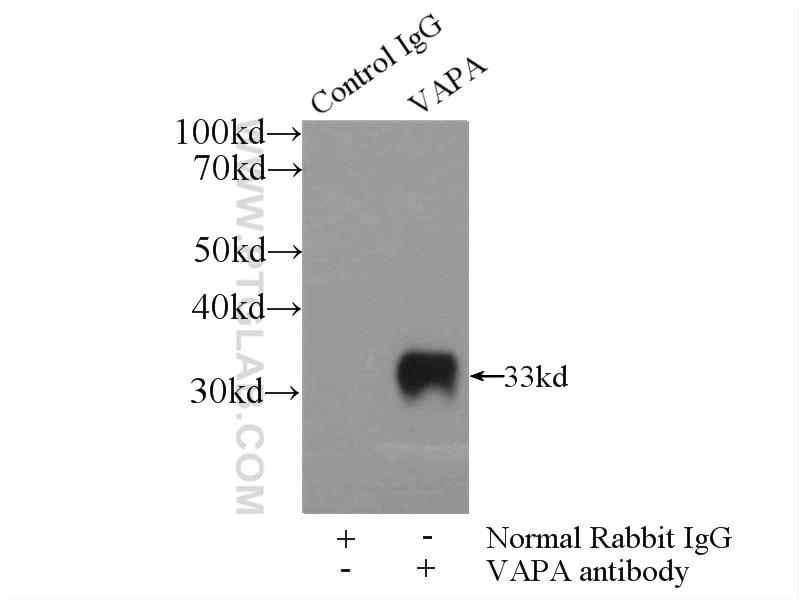
| Résultats positifs en IP | cellules HEK-293 |
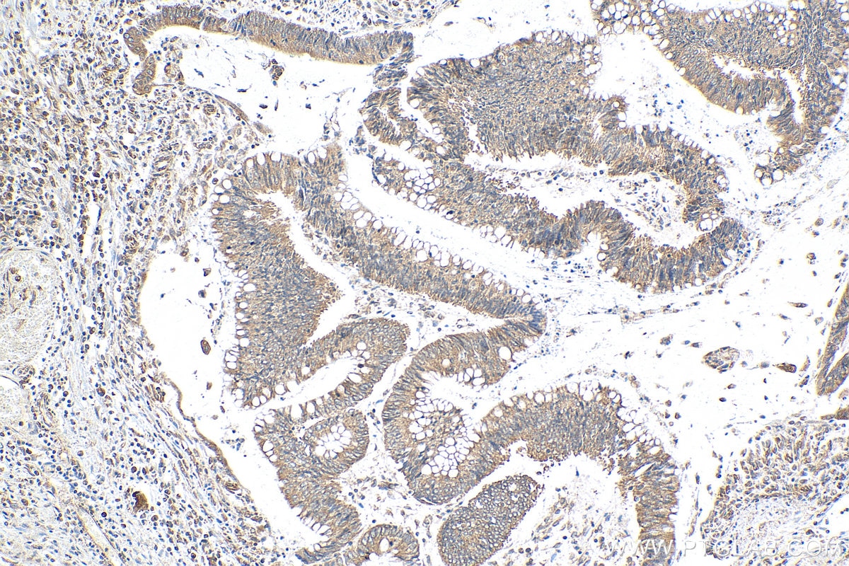
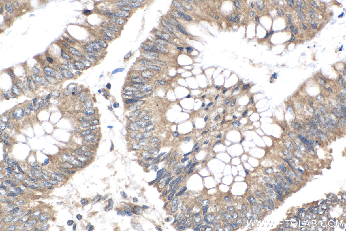
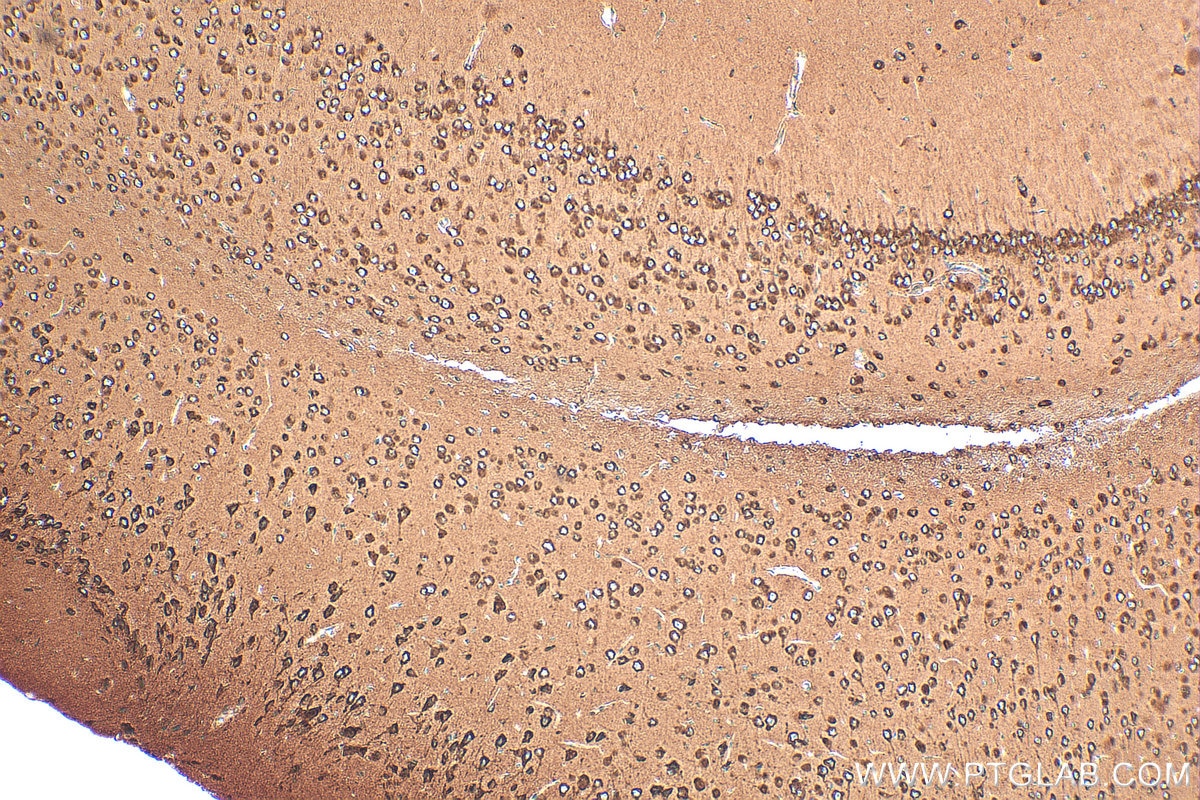
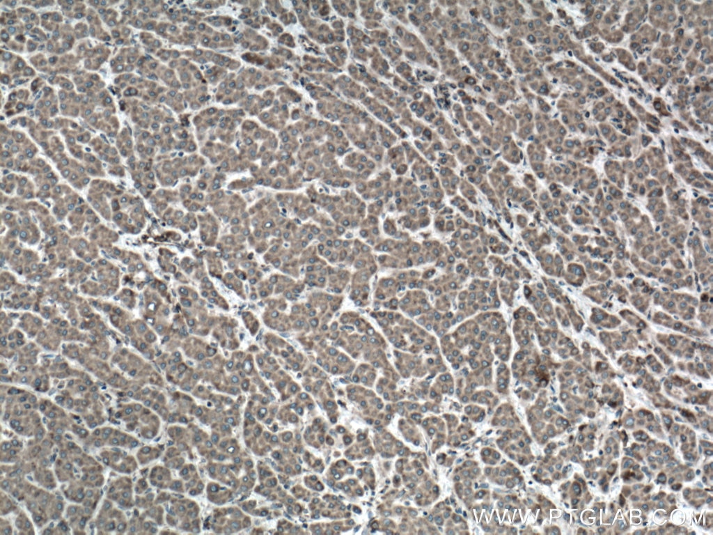
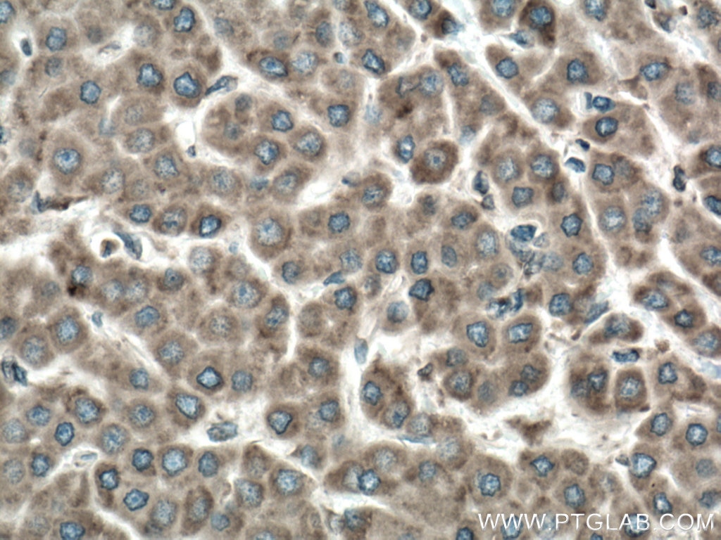
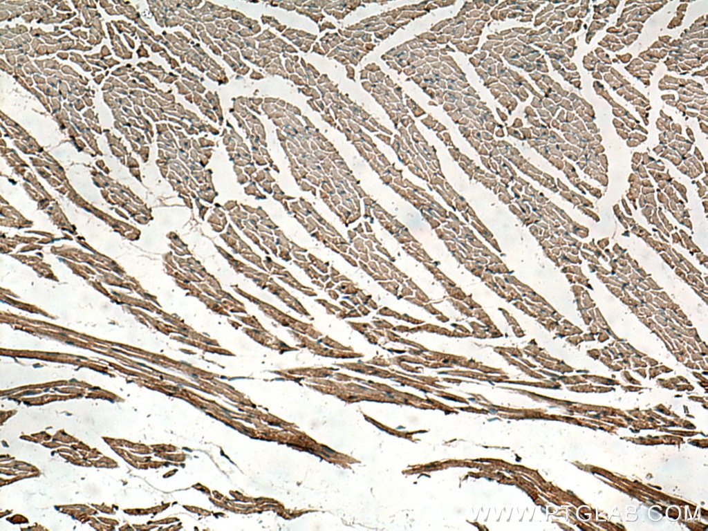
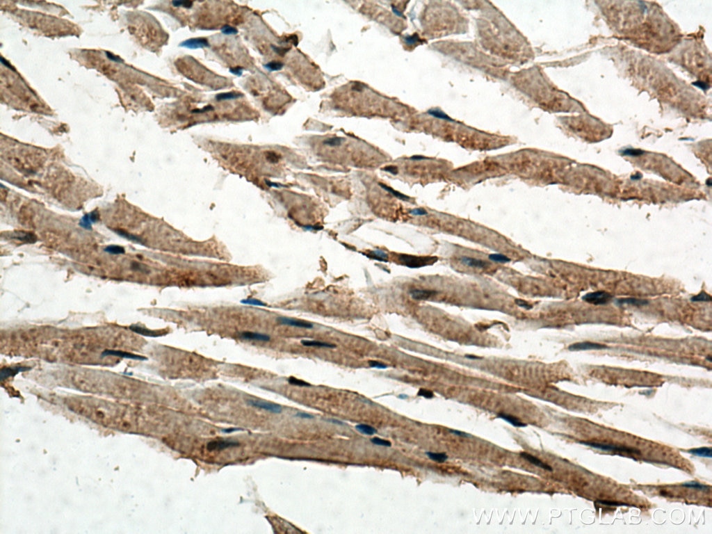
| Résultats positifs en IHC | tissu de cancer du côlon humain, tissu cardiaque de souris, tissu cérébral de souris, tissu de cancer du foie humain il est suggéré de démasquer l'antigène avec un tampon de TE buffer pH 9.0; (*) À défaut, 'le démasquage de l'antigène peut être 'effectué avec un tampon citrate pH 6,0. |
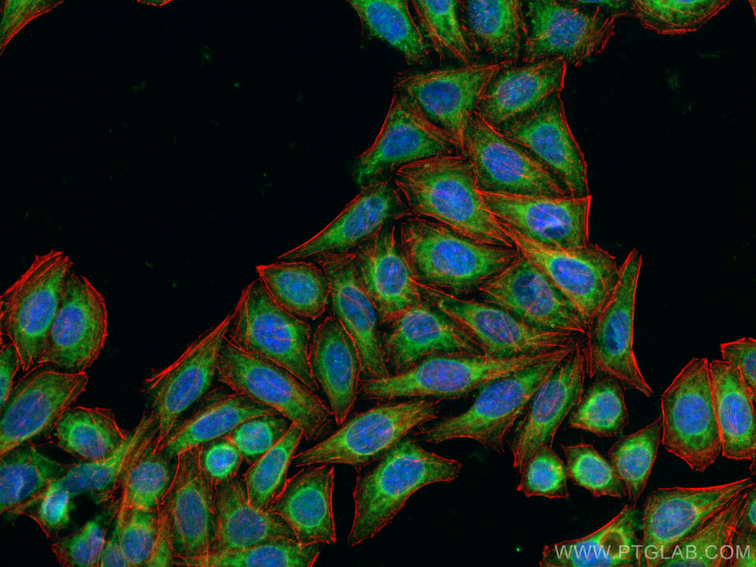
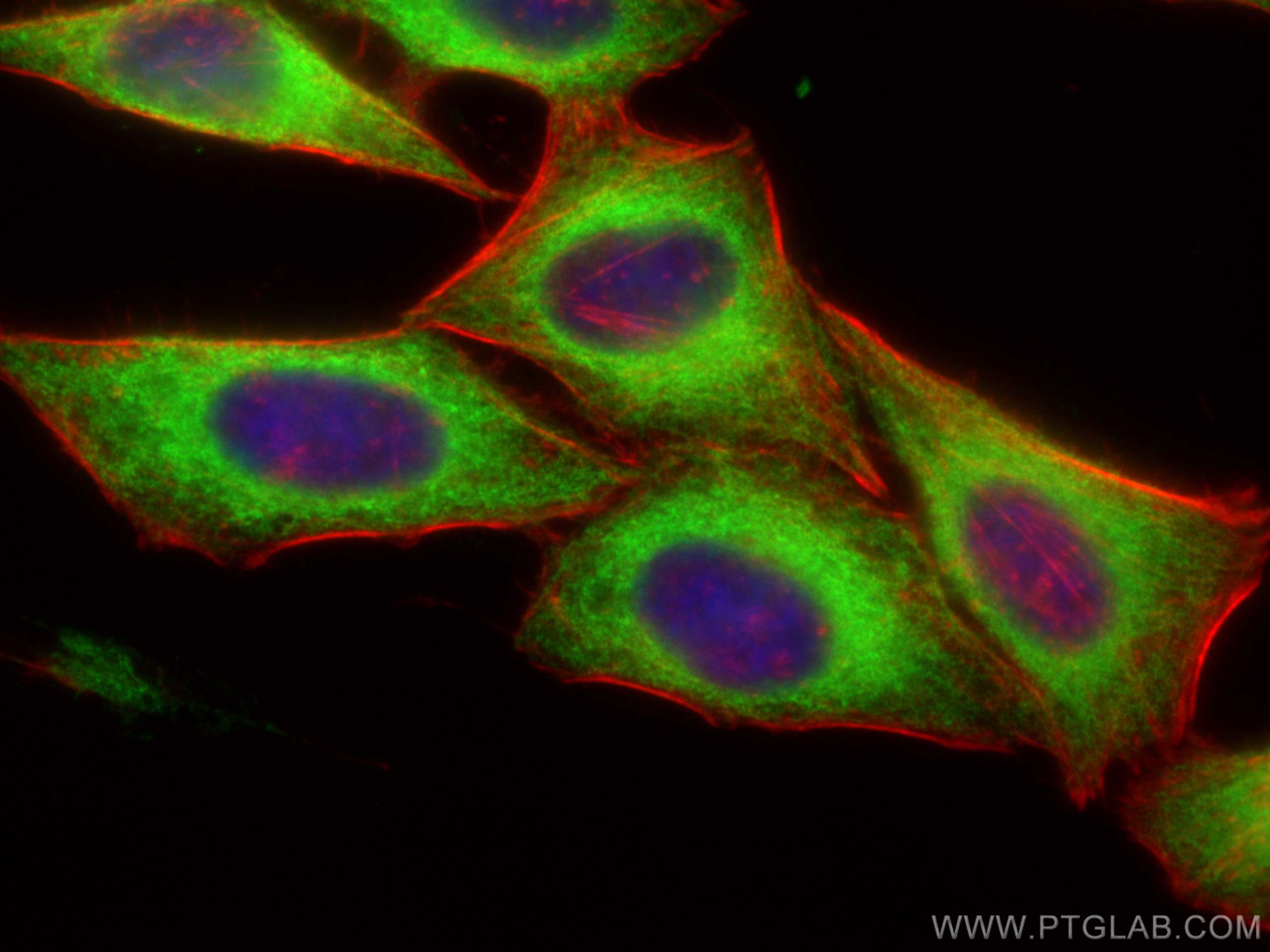
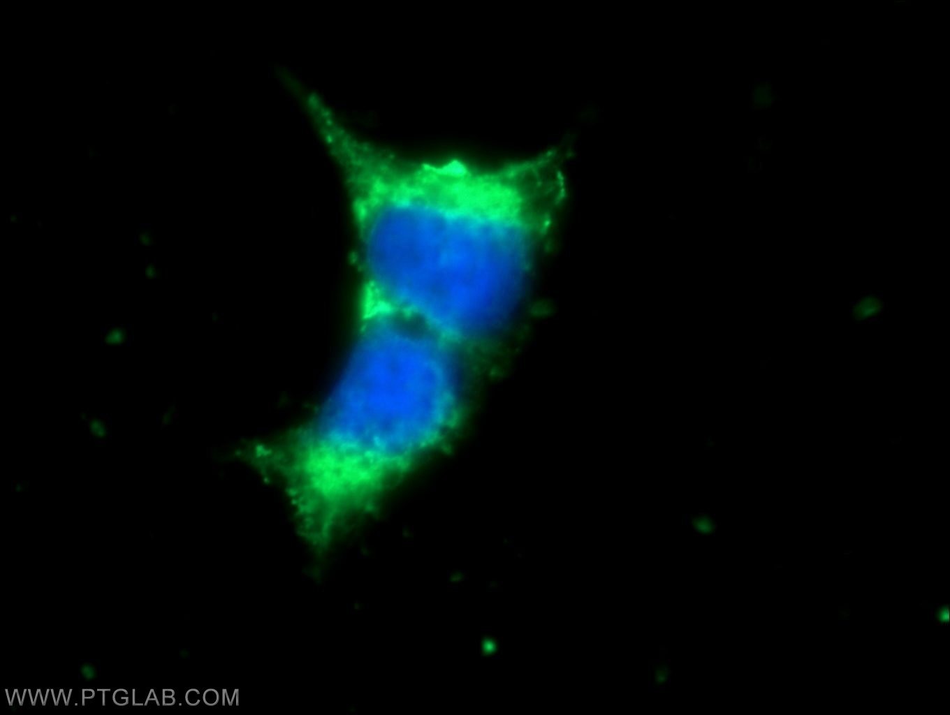
| Résultats positifs en IF/ICC | cellules HepG2, cellules HEK-293 |
Dilution recommandée
| Application | Dilution |
|---|---|
| Western Blot (WB) | WB : 1:2000-1:16000 |
| Immunoprécipitation (IP) | IP : 0.5-4.0 ug for 1.0-3.0 mg of total protein lysate |
| Immunohistochimie (IHC) | IHC : 1:200-1:1600 |
| Immunofluorescence (IF)/ICC | IF/ICC : 1:200-1:800 |
| It is recommended that this reagent should be titrated in each testing system to obtain optimal results. | |
| Sample-dependent, check data in validation data gallery | |
Applications publiées
| KD/KO | See 3 publications below |
| WB | See 21 publications below |
| IHC | See 1 publications below |
| IF | See 11 publications below |
| CoIP | See 3 publications below |
Informations sur le produit
15275-1-AP cible VAPA dans les applications de WB, IHC, IF/ICC, IP, COIP, ELISA et montre une réactivité avec des échantillons Humain, rat, souris
| Réactivité | Humain, rat, souris |
| Réactivité citée | rat, Humain, singe, souris |
| Hôte / Isotype | Lapin / IgG |
| Clonalité | Polyclonal |
| Type | Anticorps |
| Immunogène | VAPA Protéine recombinante Ag7393 |
| Nom complet | VAMP (vesicle-associated membrane protein)-associated protein A, 33kDa |
| Masse moléculaire calculée | 28 kDa |
| Poids moléculaire observé | 28-30 kDa |
| Numéro d’acquisition GenBank | BC002992 |
| Symbole du gène | VAPA |
| Identification du gène (NCBI) | 9218 |
| Conjugaison | Non conjugué |
| Forme | Liquide |
| Méthode de purification | Purification par affinité contre l'antigène |
| Tampon de stockage | PBS with 0.02% sodium azide and 50% glycerol |
| Conditions de stockage | Stocker à -20°C. Stable pendant un an après l'expédition. L'aliquotage n'est pas nécessaire pour le stockage à -20oC Les 20ul contiennent 0,1% de BSA. |
Informations générales
VAPA (Vesicle-associated membrane protein-associated protein A), also known as VAP33, is a ubiquitously expressed integral membrane protein of the VAMP-associated protein (VAP) family. It is present in the plasma membrane and in intracellular vesicles, playing a role in vesicle trafficking.
Protocole
| Product Specific Protocols | |
|---|---|
| WB protocol for VAPA antibody 15275-1-AP | Download protocol |
| IHC protocol for VAPA antibody 15275-1-AP | Download protocol |
| IF protocol for VAPA antibody 15275-1-AP | Download protocol |
| IP protocol for VAPA antibody 15275-1-AP | Download protocol |
| Standard Protocols | |
|---|---|
| Click here to view our Standard Protocols |
Publications
| Species | Application | Title |
|---|---|---|
Mol Cell The ER-Localized Transmembrane Protein EPG-3/VMP1 Regulates SERCA Activity to Control ER-Isolation Membrane Contacts for Autophagosome Formation. | ||
Mol Cell MIGA2 Links Mitochondria, the ER, and Lipid Droplets and Promotes De Novo Lipogenesis in Adipocytes. | ||
J Clin Invest Upregulation of Rubicon promotes autosis during myocardial ischemia/reperfusion injury. | ||
EMBO J A selective ER-phagy exerts procollagen quality control via a Calnexin-FAM134B complex. | ||
Curr Biol The ER Contact Proteins VAPA/B Interact with Multiple Autophagy Proteins to Modulate Autophagosome Biogenesis.
|
Avis
The reviews below have been submitted by verified Proteintech customers who received an incentive for providing their feedback.
FH Janathan (Verified Customer) (12-21-2023) | Really good and strong signal in WB and IF. IP works on WB, but not sensitive enough for visualizing with coomassie
|
FH Wei (Verified Customer) (09-22-2020) | The antibody have high specific band in western blot, but not so good for heart sections.
|
