- Phare
- Validé par KD/KO
Anticorps Polyclonal de lapin anti-VAPB
VAPB Polyclonal Antibody for WB, IHC, IF/ICC, FC (Intra), IP, ELISA
Hôte / Isotype
Lapin / IgG
Réactivité testée
Humain, rat, souris et plus (2)
Applications
WB, IHC, IF/ICC, FC (Intra), IP, CoIP, ELISA
Conjugaison
Non conjugué
N° de cat : 14477-1-AP
Synonymes
Galerie de données de validation
Applications testées
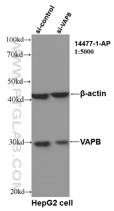
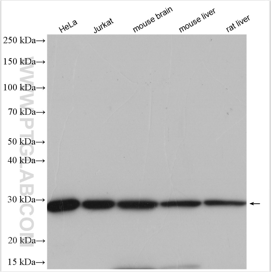
| Résultats positifs en WB | cellules HeLa, cellules HepG2, cellules Jurkat, tissu cérébral de souris, tissu hépatique de rat, tissu hépatique de souris |
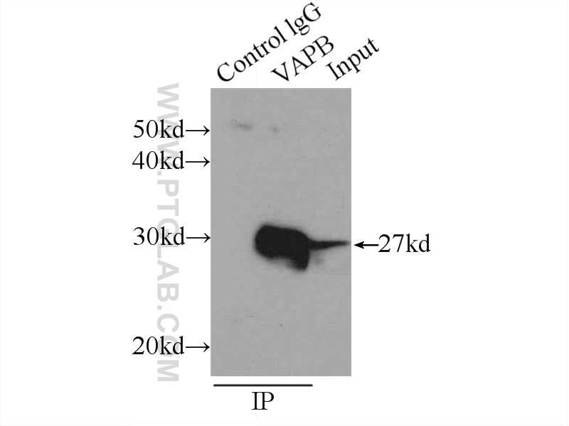
| Résultats positifs en IP | cellules HeLa |
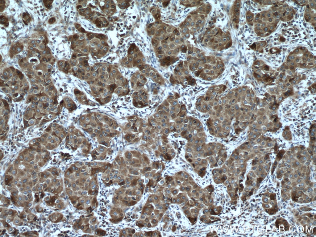
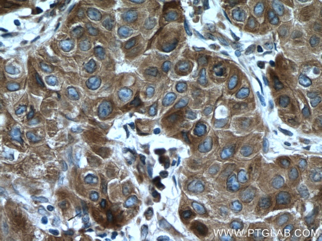
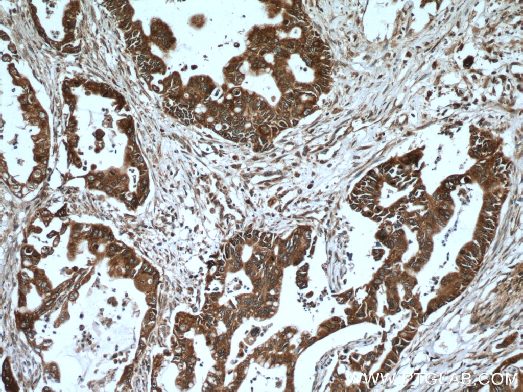
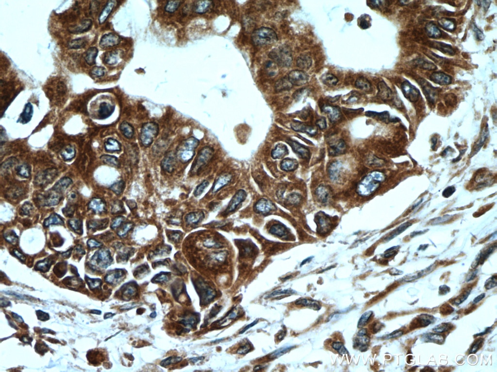
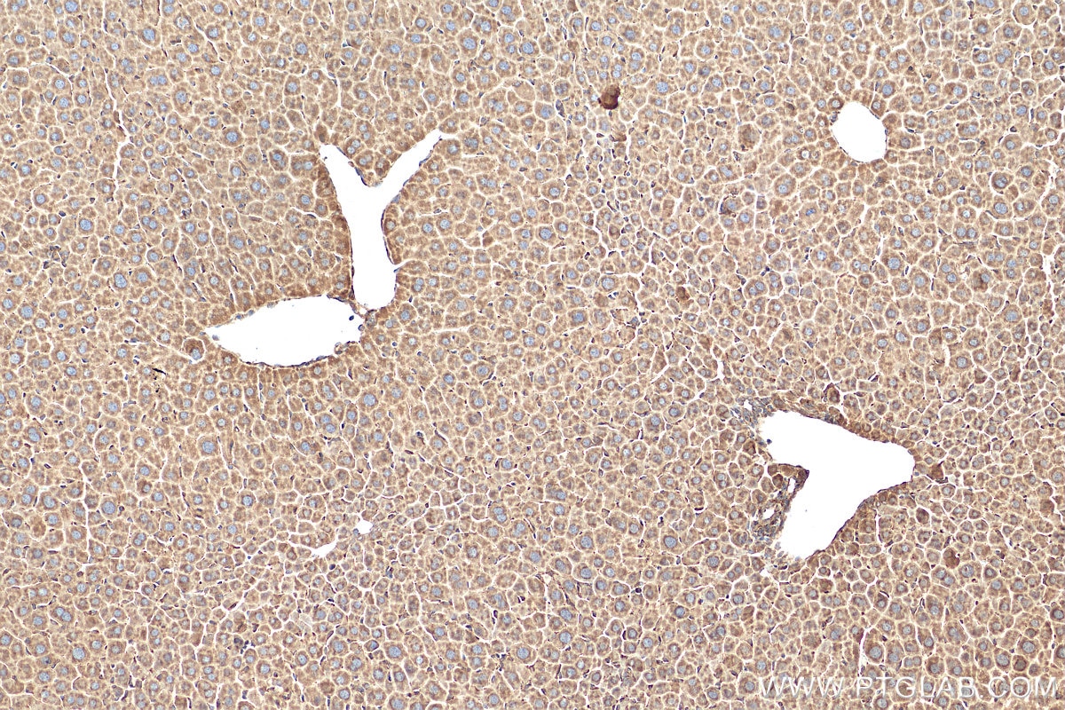
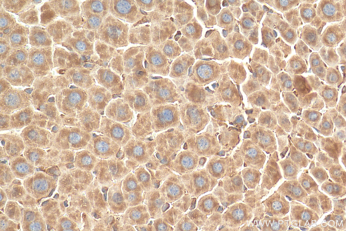
| Résultats positifs en IHC | tissu de cancer du sein humain, tissu de cancer du pancréas humain, tissu hépatique de souris il est suggéré de démasquer l'antigène avec un tampon de TE buffer pH 9.0; (*) À défaut, 'le démasquage de l'antigène peut être 'effectué avec un tampon citrate pH 6,0. |
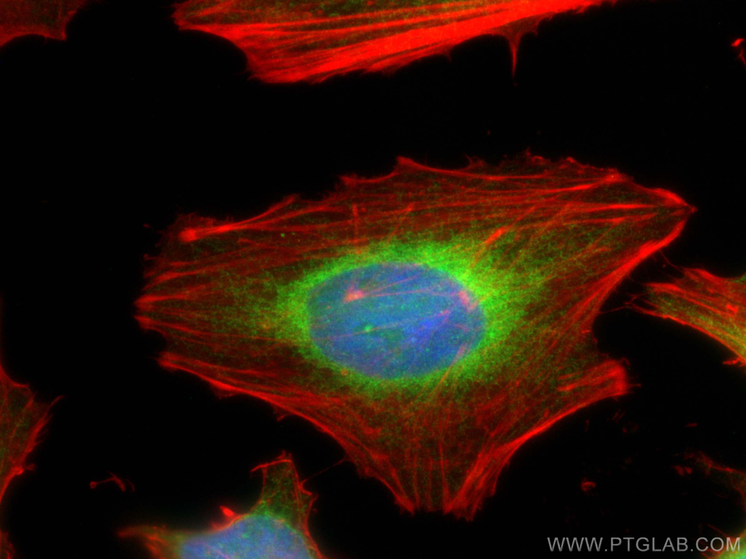
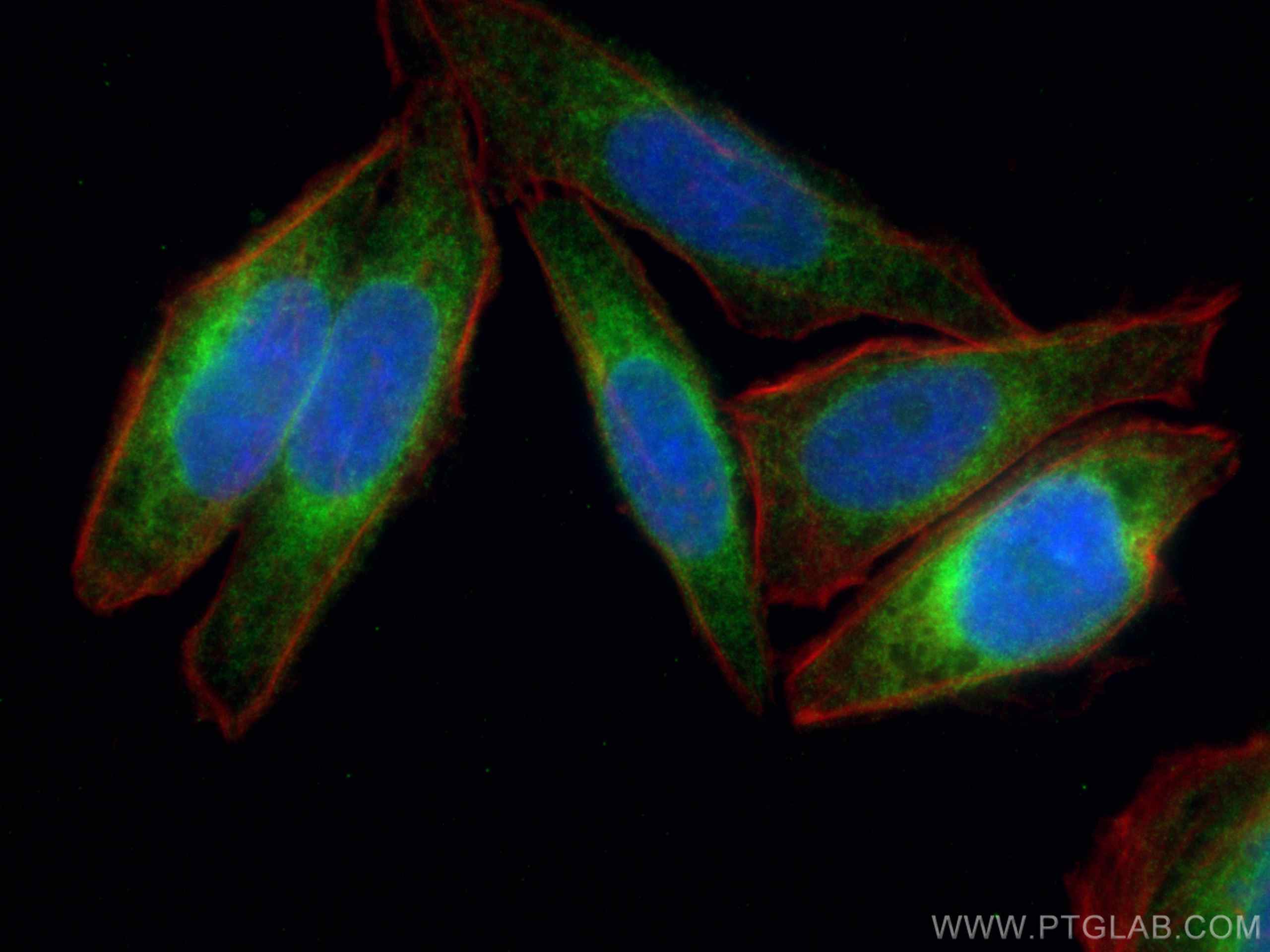
| Résultats positifs en IF/ICC | cellules HeLa, cellules HepG2 |
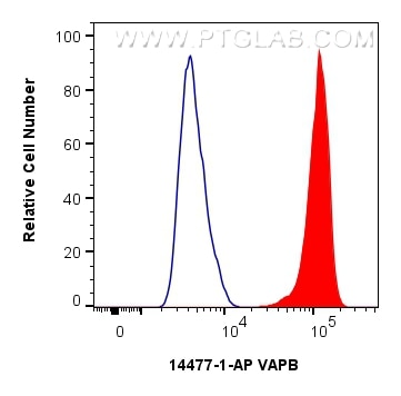
| Résultats positifs en FC (Intra) | cellules HepG2, |
Dilution recommandée
| Application | Dilution |
|---|---|
| Western Blot (WB) | WB : 1:2000-1:12000 |
| Immunoprécipitation (IP) | IP : 0.5-4.0 ug for 1.0-3.0 mg of total protein lysate |
| Immunohistochimie (IHC) | IHC : 1:50-1:500 |
| Immunofluorescence (IF)/ICC | IF/ICC : 1:400-1:1600 |
| Flow Cytometry (FC) (INTRA) | FC (INTRA) : 0.25 ug per 10^6 cells in a 100 µl suspension |
| It is recommended that this reagent should be titrated in each testing system to obtain optimal results. | |
| Sample-dependent, check data in validation data gallery | |
Applications publiées
| KD/KO | See 4 publications below |
| WB | See 44 publications below |
| IHC | See 1 publications below |
| IF | See 16 publications below |
| IP | See 2 publications below |
| CoIP | See 2 publications below |
Informations sur le produit
14477-1-AP cible VAPB dans les applications de WB, IHC, IF/ICC, FC (Intra), IP, CoIP, ELISA et montre une réactivité avec des échantillons Humain, rat, souris
| Réactivité | Humain, rat, souris |
| Réactivité citée | rat, Humain, poisson-zèbre, singe, souris |
| Hôte / Isotype | Lapin / IgG |
| Clonalité | Polyclonal |
| Type | Anticorps |
| Immunogène | VAPB Protéine recombinante Ag5857 |
| Nom complet | VAMP (vesicle-associated membrane protein)-associated protein B and C |
| Masse moléculaire calculée | 27 kDa |
| Poids moléculaire observé | 27 kDa |
| Numéro d’acquisition GenBank | BC001712 |
| Symbole du gène | VAPB |
| Identification du gène (NCBI) | 9217 |
| Conjugaison | Non conjugué |
| Forme | Liquide |
| Méthode de purification | Purification par affinité contre l'antigène |
| Tampon de stockage | PBS with 0.02% sodium azide and 50% glycerol |
| Conditions de stockage | Stocker à -20°C. Stable pendant un an après l'expédition. L'aliquotage n'est pas nécessaire pour le stockage à -20oC Les 20ul contiennent 0,1% de BSA. |
Informations générales
Vesicle-associated membrane protein-associated protein B (VAPB) is an integral membrane protein localized to the endoplasmic reticulum (ER) membrane. VAPB has been implicated in various cellular processes, including ER stress, the unfolded protein response (UPR) and calcium homeostasis regulation. The mutations in the gene of VAPB cause amyotrophic lateral sclerosis 8 (ALS8) and some other related forms of motor neuron disease including late-onset spinal muscular atrophy.
Protocole
| Product Specific Protocols | |
|---|---|
| WB protocol for VAPB antibody 14477-1-AP | Download protocol |
| IHC protocol for VAPB antibody 14477-1-AP | Download protocol |
| IF protocol for VAPB antibody 14477-1-AP | Download protocol |
| IP protocol for VAPB antibody 14477-1-AP | Download protocol |
| Standard Protocols | |
|---|---|
| Click here to view our Standard Protocols |
Publications
| Species | Application | Title |
|---|---|---|
Nat Cell Biol MIROs and DRP1 drive mitochondrial-derived vesicle biogenesis and promote quality control. | ||
Cell Res DNA damage triggers tubular endoplasmic reticulum extension to promote apoptosis by facilitating ER-mitochondria signaling. | ||
Mol Cell MIGA2 Links Mitochondria, the ER, and Lipid Droplets and Promotes De Novo Lipogenesis in Adipocytes. | ||
J Clin Invest SPTLC1 variants associated with ALS produce distinct sphingolipid signatures through impaired interaction with ORMDL proteins. | ||
Cancer Res Proteomic mapping and targeting of mitotic pericentriolar material in tumors bearing centrosome amplification. |
Avis
The reviews below have been submitted by verified Proteintech customers who received an incentive for providing their feedback.
FH Wei (Verified Customer) (09-22-2020) | The antibody have predicted band on western blot.
|
