- Phare
- Validé par KD/KO
Anticorps Polyclonal de lapin anti-VPS26A
VPS26A Polyclonal Antibody for WB, IHC, IF/ICC, ELISA
Hôte / Isotype
Lapin / IgG
Réactivité testée
Humain, rat, souris
Applications
WB, IHC, IF/ICC, ELISA
Conjugaison
Non conjugué
N° de cat : 12804-1-AP
Synonymes
Galerie de données de validation
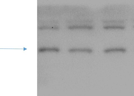
 at dilution of 1:600 incubated at room temperature for 1.5 hours.")
 at dilution of 1:300 incubated at room temperature for 1.5 hours.")
 at dilution of 1:200 (under 10x lens). Heat mediated antigen retrieval with Tris-EDTA buffer (pH 9.0).")
 at dilution of 1:200 (under 40x lens). Heat mediated antigen retrieval with Tris-EDTA buffer (pH 9.0).")
 at dilution of 1:100 (under 10x lens).")
 at dilution of 1:200 (under 10x lens).")
 at dilution of 1:200 (under 40x lens).")
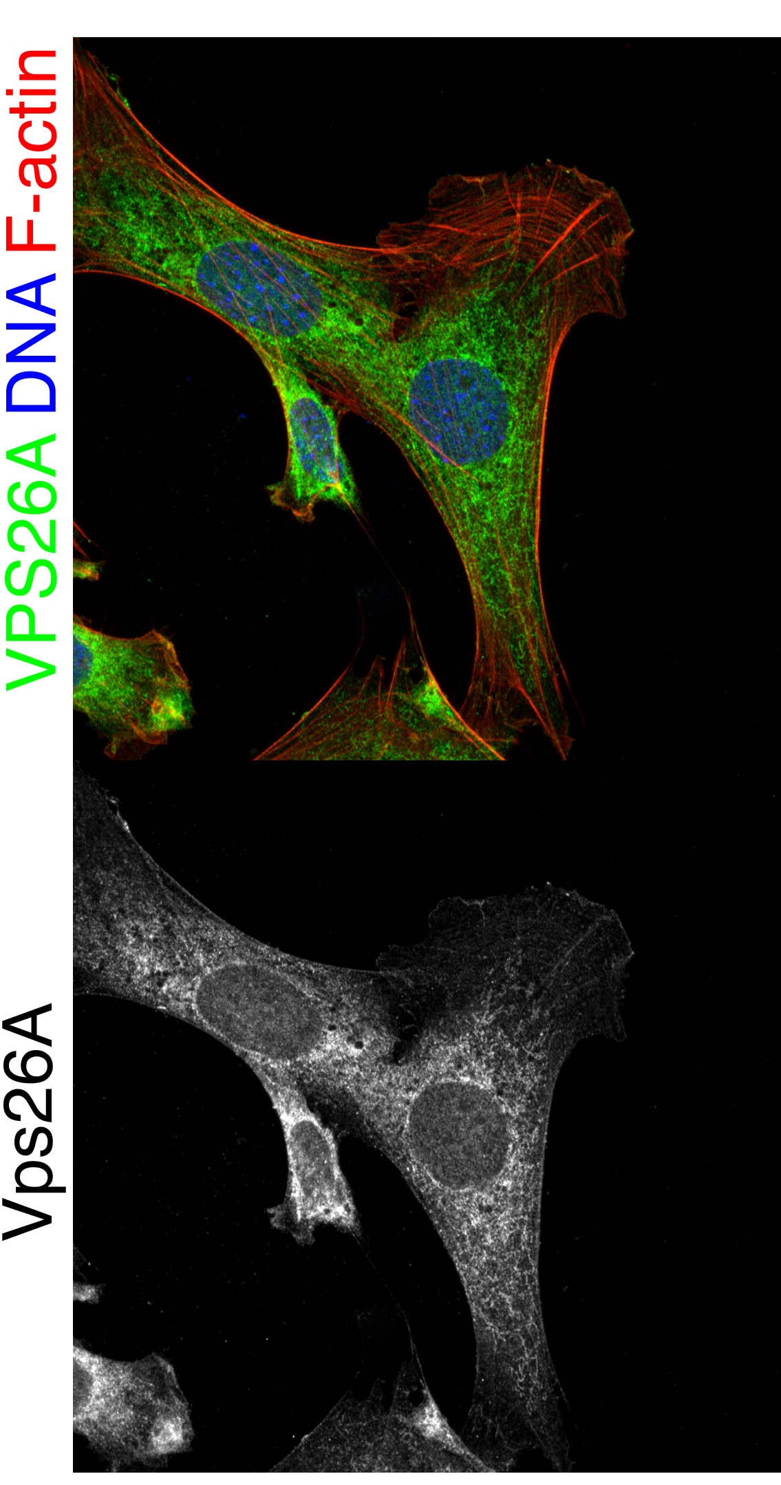
 fixed HeLa cells using 12804-1-AP (VPS26A antibody), at dilution of 1:200 and CoraLite®488-Conjugated Goat Anti-Rabbit IgG(H+L), CL555-phalloidine stains F-actin (red).")
 at dilution of 1:50 and Alexa Fluor 488-conjugated Goat Anti-Rabbit IgG(H+L).")
Applications testées
| Résultats positifs en WB | tissu rénal de souris, cellules HEK-293, tissu rénal de rat |
| Résultats positifs en IHC | tissu de cancer du sein humain, tissu de cancer du col de l'utérus humain, tissu rénal humain il est suggéré de démasquer l'antigène avec un tampon de TE buffer pH 9.0; (*) À défaut, 'le démasquage de l'antigène peut être 'effectué avec un tampon citrate pH 6,0. |
| Résultats positifs en IF/ICC | cellules HeLa, |
Dilution recommandée
| Application | Dilution |
|---|---|
| Western Blot (WB) | WB : 1:500-1:1000 |
| Immunohistochimie (IHC) | IHC : 1:50-1:500 |
| Immunofluorescence (IF)/ICC | IF/ICC : 1:50-1:500 |
| It is recommended that this reagent should be titrated in each testing system to obtain optimal results. | |
| Sample-dependent, check data in validation data gallery | |
Applications publiées
| KD/KO | See 1 publications below |
| WB | See 7 publications below |
| IHC | See 1 publications below |
| IF | See 2 publications below |
Informations sur le produit
12804-1-AP cible VPS26A dans les applications de WB, IHC, IF/ICC, ELISA et montre une réactivité avec des échantillons Humain, rat, souris
| Réactivité | Humain, rat, souris |
| Réactivité citée | Humain |
| Hôte / Isotype | Lapin / IgG |
| Clonalité | Polyclonal |
| Type | Anticorps |
| Immunogène | VPS26A Protéine recombinante Ag3391 |
| Nom complet | vacuolar protein sorting 26 homolog A (S. pombe) |
| Masse moléculaire calculée | 38 kDa |
| Poids moléculaire observé | 38 kDa |
| Numéro d’acquisition GenBank | BC022505 |
| Symbole du gène | VPS26A |
| Identification du gène (NCBI) | 9559 |
| Conjugaison | Non conjugué |
| Forme | Liquide |
| Méthode de purification | Purification par affinité contre l'antigène |
| Tampon de stockage | PBS with 0.02% sodium azide and 50% glycerol |
| Conditions de stockage | Stocker à -20°C. Stable pendant un an après l'expédition. L'aliquotage n'est pas nécessaire pour le stockage à -20oC Les 20ul contiennent 0,1% de BSA. |
Informations générales
In mammals, there are two paralogues of yeast Vps26, VPS26A and VPS26B (PMID: 16190980). VPS26 is a component of the retromer complex composed of VPS26 (VPS26A or VPS26B), VPS29, VPS35, SNX1, and SNX2. VPS26A and VPS26B subunits define distinct retromer complexes (PMID: 21920005). The retromer complex is important in recycling transmembrane receptors from endosomes to the trans-Golgi network (TGN).
Protocole
| Product Specific Protocols | |
|---|---|
| WB protocol for VPS26A antibody 12804-1-AP | Download protocol |
| IHC protocol for VPS26A antibody 12804-1-AP | Download protocol |
| IF protocol for VPS26A antibody 12804-1-AP | Download protocol |
| Standard Protocols | |
|---|---|
| Click here to view our Standard Protocols |
Publications
| Species | Application | Title |
|---|---|---|
Cell Metab Phospholipase PLA2G6, a Parkinsonism-Associated Gene, Affects Vps26 and Vps35, Retromer Function, and Ceramide Levels, Similar to α-Synuclein Gain. | ||
bioRxiv Noncanonical roles of ATG5 and membrane atg8ylation in retromer assembly and function | ||
Elife Noncanonical roles of ATG5 and membrane atg8ylation in retromer assembly and function | ||
Autophagy RAB21 controls autophagy and cellular energy homeostasis by regulating retromer-mediated recycling of SLC2A1/GLUT1 | ||
Int J Mol Sci The Prognostic Value and the Oncogenic and Immunological Roles of Vacuolar Protein Sorting Associated Protein 26 A in Pancreatic Adenocarcinoma
|
Avis
The reviews below have been submitted by verified Proteintech customers who received an incentive for providing their feedback.
FH Xin (Verified Customer) (10-10-2022) | It is Ok to detect endogenous VPS26A although there are some non-specific bands.
 |
FH Stephen (Verified Customer) (09-07-2019) | Oc-2 cells fixed with 4% PFA Perm. by 0.3% tx100 for 5 min blocked with 1% BSA in 1XPBS for 2 hours vps26a antibody incubated 1:300 overnight at 4 degrees in 1%BSA in 1x PBS. Co-stained with DAPI (blue) and Ph647 (red) to visualize DNA and F-actin respectively.
 |

