- Phare
- Validé par KD/KO
Anticorps Polyclonal de lapin anti-VTI1B
VTI1B Polyclonal Antibody for WB, IP, IHC, ELISA
Hôte / Isotype
Lapin / IgG
Réactivité testée
Humain, rat, souris
Applications
WB, IP, IF, IHC, ELISA
Conjugaison
Non conjugué
N° de cat : 14495-1-AP
Synonymes
Galerie de données de validation
Applications testées
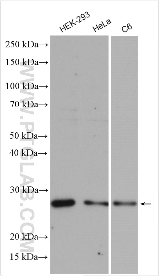
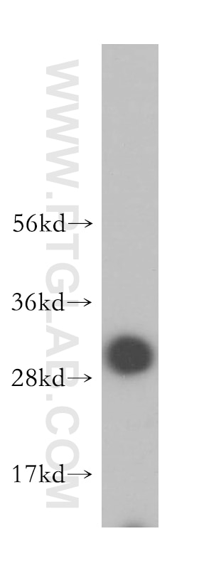
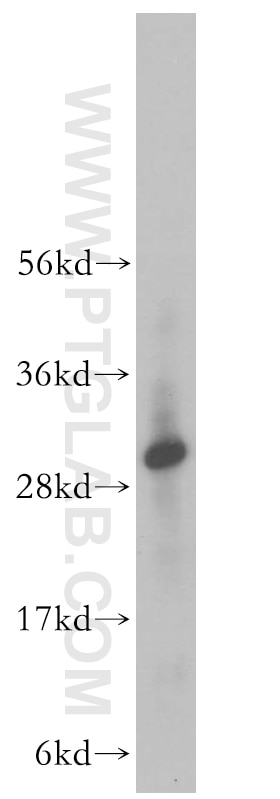
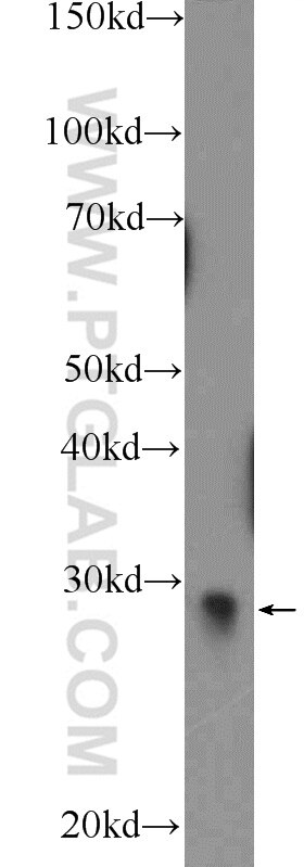
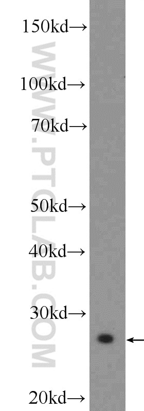
| Résultats positifs en WB | cellules HEK-293, cellules C6, cellules HeLa, cellules NIH/3T3, tissu hépatique humain |
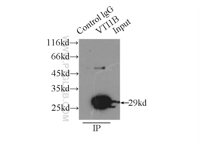
| Résultats positifs en IP | cellules HeLa |
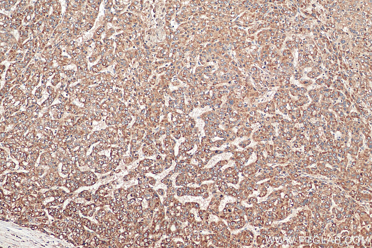
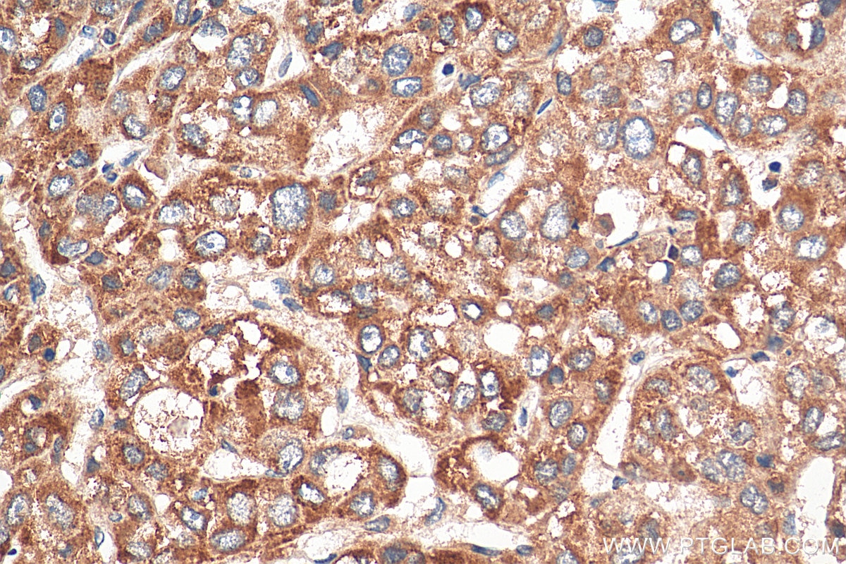
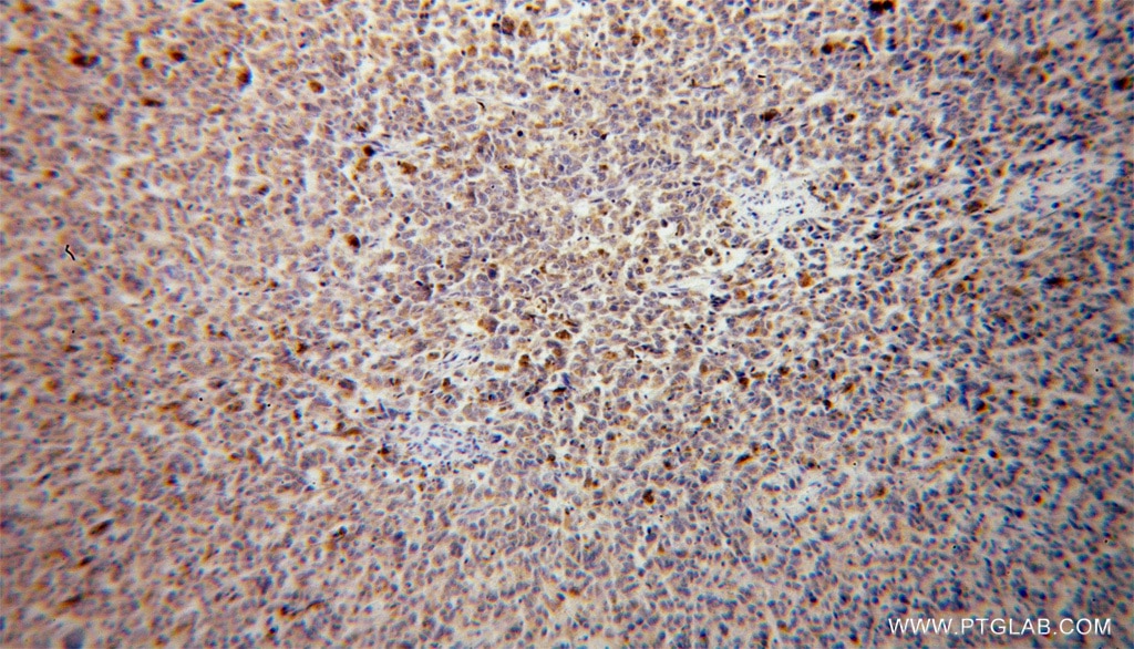
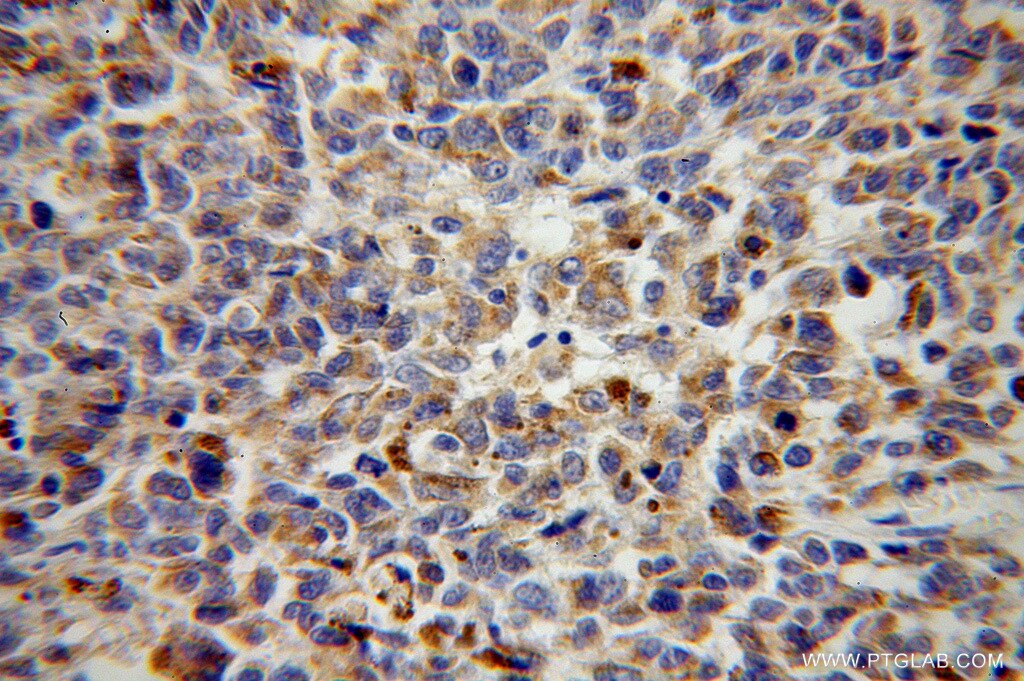
| Résultats positifs en IHC | tissu de cancer du foie humain, tissu de mélanome malin humain il est suggéré de démasquer l'antigène avec un tampon de TE buffer pH 9.0; (*) À défaut, 'le démasquage de l'antigène peut être 'effectué avec un tampon citrate pH 6,0. |
Dilution recommandée
| Application | Dilution |
|---|---|
| Western Blot (WB) | WB : 1:1000-1:4000 |
| Immunoprécipitation (IP) | IP : 0.5-4.0 ug for 1.0-3.0 mg of total protein lysate |
| Immunohistochimie (IHC) | IHC : 1:50-1:500 |
| It is recommended that this reagent should be titrated in each testing system to obtain optimal results. | |
| Sample-dependent, check data in validation data gallery | |
Applications publiées
| KD/KO | See 1 publications below |
| WB | See 2 publications below |
| IF | See 1 publications below |
Informations sur le produit
14495-1-AP cible VTI1B dans les applications de WB, IP, IF, IHC, ELISA et montre une réactivité avec des échantillons Humain, rat, souris
| Réactivité | Humain, rat, souris |
| Réactivité citée | Humain, souris |
| Hôte / Isotype | Lapin / IgG |
| Clonalité | Polyclonal |
| Type | Anticorps |
| Immunogène | VTI1B Protéine recombinante Ag5906 |
| Nom complet | vesicle transport through interaction with t-SNAREs homolog 1B (yeast) |
| Masse moléculaire calculée | 27 kDa |
| Poids moléculaire observé | 29 kDa |
| Numéro d’acquisition GenBank | BC003142 |
| Symbole du gène | VTI1B |
| Identification du gène (NCBI) | 10490 |
| Conjugaison | Non conjugué |
| Forme | Liquide |
| Méthode de purification | Purification par affinité contre l'antigène |
| Tampon de stockage | PBS with 0.02% sodium azide and 50% glycerol |
| Conditions de stockage | Stocker à -20°C. Stable pendant un an après l'expédition. L'aliquotage n'est pas nécessaire pour le stockage à -20oC Les 20ul contiennent 0,1% de BSA. |
Informations générales
Fusion between membranes is mediated by specific SNARE (soluble N-ethylmeleimide-sensitive factor attachment protein receptor) complexes. Two human SNARE proteins, VTI1A and VTI1B, are homologous to the yeast Q-SNARE Vtilp which is part of several SNARE complexes in different transport steps (PMID: 12067063). Both proteins had a distinct but overlapping localization. VTI1A is localized predominantly in the TGN, VTI1B in late endosomes (PMID:12067063; 21262811). VTI1B forms a SNARE complex with STX7, STX8 and VAMP8 which functions in the homotypic fusion of late endosomes. It is a component of the SNARE complex composed of STX7, STX8, VAMP7 and VIT1B that is required for heterotypic fusion of late endosomes with lysosomes. It has also been reported that VIT1B interacts with EpsinR, a protein involved in exocytic trafficking (PMID: 15371541).
Protocole
| Product Specific Protocols | |
|---|---|
| WB protocol for VTI1B antibody 14495-1-AP | Download protocol |
| IHC protocol for VTI1B antibody 14495-1-AP | Download protocol |
| IP protocol for VTI1B antibody 14495-1-AP | Download protocol |
| Standard Protocols | |
|---|---|
| Click here to view our Standard Protocols |
Publications
| Species | Application | Title |
|---|---|---|
Autophagy The STX6-VTI1B-VAMP3 complex facilitates xenophagy by regulating the fusion between recycling endosomes and autophagosomes.
| ||
mBio Syntaxin 11 Contributes to the Interferon-Inducible Restriction of Coxiella burnetii Intracellular Infection | ||
Front Cell Dev Biol The SNARE protein Vti1b is recruited to the sites of BCR activation but is redundant for antigen internalisation, processing and presentation |
Avis
The reviews below have been submitted by verified Proteintech customers who received an incentive for providing their feedback.
FH Tongbin (Verified Customer) (08-25-2020) | This antibody had a specific band at VTI1B's predicted MW ~28 kDa.
|
